Articles | Volume 34, issue 6
https://doi.org/10.5194/ejm-34-573-2022
© Author(s) 2022. This work is distributed under
the Creative Commons Attribution 4.0 License.
the Creative Commons Attribution 4.0 License.
https://doi.org/10.5194/ejm-34-573-2022
© Author(s) 2022. This work is distributed under
the Creative Commons Attribution 4.0 License.
the Creative Commons Attribution 4.0 License.
Research article
| 
09 Dec 2022
Research article |
| 09 Dec 2022
| 09 Dec 2022
Non-destructive determination of the biotite crystal chemistry using Raman spectroscopy: how far we can go?
Stylianos Aspiotis
CORRESPONDING AUTHOR
Fachbereich Erdsystemwissenschaften, Universität Hamburg,
Grindelallee 48, 20146 Hamburg, Germany
Jochen Schlüter
Museum der Natur Hamburg – Mineralogie, Leibniz-Institut zur Analyse des
Biodiversitätswandels (LIB), Grindelallee 48, 20146 Hamburg, Germany
Günther J. Redhammer
Fachbereich Chemie und Physik der Materialien, Paris-Lodron
Universität Salzburg, 5020 Salzburg, Austria
Boriana Mihailova
Fachbereich Erdsystemwissenschaften, Universität Hamburg,
Grindelallee 48, 20146 Hamburg, Germany
Related authors
Christian Reinberg, Stylianos Aspiotis, Thomas Malcherek, Stefan T. M. Peters, and Boriana Mihailova
Eur. J. Mineral., 38, 461–475, https://doi.org/10.5194/ejm-38-461-2026, https://doi.org/10.5194/ejm-38-461-2026, 2026
Short summary
Short summary
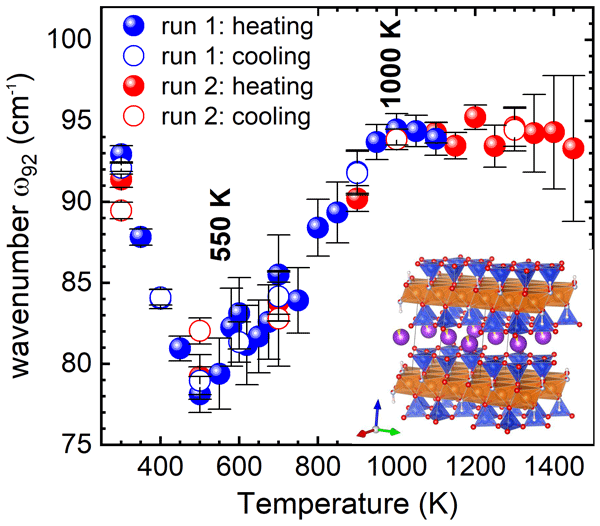
By applying in situ high-temperature Raman spectroscopy to (OH)-- and Fe2+-bearing fluorophlogopite we show that a phonon-driven structural instability near 600 K triggers the mobilization of interlayer K+ cations, which can act as charge carriers. Above 1100 K all H+ cations delocalize and can also contribute to transport processes. Above 1300 K oxidation of Fe takes place, along with limited dehydrogenation and dehydroxylation, facilitating partial K+ leakage and structural decomposition.
Christian Reinberg, Stylianos Aspiotis, Thomas Malcherek, Stefan T. M. Peters, and Boriana Mihailova
Eur. J. Mineral., 38, 461–475, https://doi.org/10.5194/ejm-38-461-2026, https://doi.org/10.5194/ejm-38-461-2026, 2026
Short summary
Short summary
By applying in situ high-temperature Raman spectroscopy to (OH)-- and Fe2+-bearing fluorophlogopite we show that a phonon-driven structural instability near 600 K triggers the mobilization of interlayer K+ cations, which can act as charge carriers. Above 1100 K all H+ cations delocalize and can also contribute to transport processes. Above 1300 K oxidation of Fe takes place, along with limited dehydrogenation and dehydroxylation, facilitating partial K+ leakage and structural decomposition.
Giancarlo Della Ventura, Roberta Oberti, Valeria Misiti, Francesco Radica, Gunther J. Redhammer, Simone Bernardini, Massimo Boiocchi, and Boriana Mihailova
Eur. J. Mineral., 37, 535–547, https://doi.org/10.5194/ejm-37-535-2025, https://doi.org/10.5194/ejm-37-535-2025, 2025
Short summary
Short summary
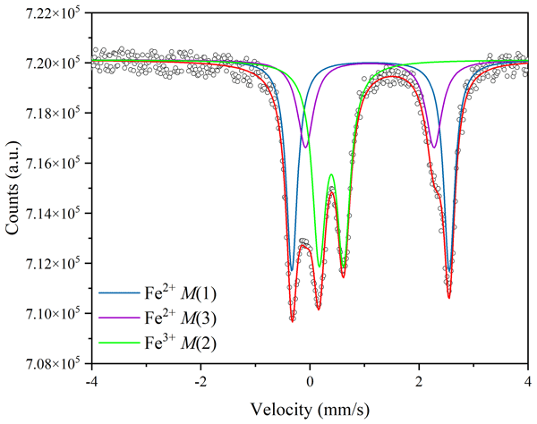
The thermal reaction Fe2+ + OH- → Fe3+ + O2- + e- + H+ in hydrous silicates has tremendous implications in planetary-scale phenomena like electrical anomalies, water cycling, seismicity, volcanism, and ore generation. We annealed riebeckite crystals up to 750 °C and P = 0.7 GPa and show that the amphibole stability is extended without any Fe oxidation, implying that thermally activated electron hopping contributes to the electrical conductivity of lithospheric rocks during subduction.
Thomas Malcherek, Boriana Mihailova, Jochen Schlüter, Philippe Roth, and Nicolas Meisser
Eur. J. Mineral., 36, 153–164, https://doi.org/10.5194/ejm-36-153-2024, https://doi.org/10.5194/ejm-36-153-2024, 2024
Short summary
Short summary
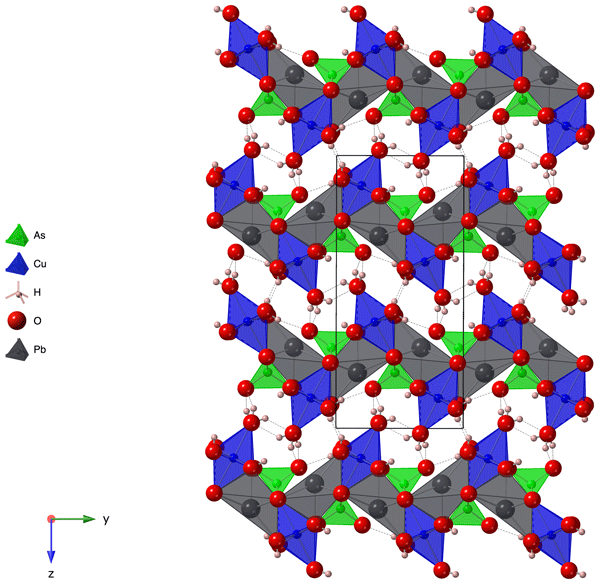
The new mineral heimite was originally discovered on the mine dumps of the Grosses Chalttal deposit, Mürtschenalp district, Glarus, Switzerland. Its relatively simple chemistry is formed by water and ions of lead, copper, arsenic, hydrogen and oxygen. The mineral's crystal structure is related to the well-known duftite, which is also observed to grow on crystals of heimite. While heimite has so far only been found in the central Alps, it is expected to occur in other copper deposits worldwide.
Mariko Nagashima and Boriana Mihailova
Eur. J. Mineral., 35, 267–283, https://doi.org/10.5194/ejm-35-267-2023, https://doi.org/10.5194/ejm-35-267-2023, 2023
Short summary
Short summary
We provide a tool for fast preparation-free estimation of the Fe3+ content in Al–Fe3+ series epidotes by Raman spectroscopy. The peaks near 570, 600, and 1090 cm−1, originating from Si2O7 vibrations, strongly correlated with Fe content, and all three signals are well resolved in a random orientation. Among them, the 570 cm−1 peak is the sharpest and easily recognized. Hence, the linear trend, ω570 = 577.1(3) − 12.7(4)x, gives highly reliable Fe content, x, with accuracy ± 0.04 Fe3+ apfu.
Cited articles
Bendel, V. and Schmidt, B. C.: Raman spectroscopic characterisation of
disordered alkali feldspars along the join
KAlSi3O8-NaAlSi3O8: application to natural sanidine and
anorthoclase, Eur. J. Mineral., 20, 1055–1065, https://doi.org/10.1127/0935-1221/2009/0021-1856, 2008.
Bersani, D. and Lottici, P. P.: Raman spectroscopy of minerals and mineral
pigments in archaeometry, J. Raman Spectrosc., 47, 499–530, 2016.
Brigatti, M. F., Frigieri, P., Ghezzo, C., and Poppi, L.: Crystal chemistry of
Al-rich biotites coexisting with muscovites in peraluminous granites, Am.
Mineral., 85, 436–448, 2000.
Brigatti, M. F., Kile, D. E., and Poppi, M.: Crystal structure and crystal
chemistry of Lithium-bearing muscovite-2M1, Can. Mineral., 39,
1171–1180, 2001.
Brigatti, M. F., Affronte, M., Elmi, C., Malferrari, D., and Laurora, A.:
Trioctahedral Fe-rich micas: Relationships between magnetic behavior and
crystal chemistry, Am. Mineral., 100, 2231–2241, 2015.
Chang, I. F. and Mitra, S. S.: Long wavelength optical phonons in mixed
crystals, Adv. Phys., 20, 359–404, 1971.
Corradini, M., de Ferri, L., and Pojana, G.: Spectroscopic characterization
of commercial pigments for pictorial retouching, J. Raman Spectrosc., 52,
35–58, 2021.
El Halim, M., Daoudi, L., El Ouahabi, M., Rousseau, V., Cools, C., and
Fagel, N.: Mineralogical and geochemical characterization of archaeological
ceramics from the 16th century El Badi Palace, Morocco, Clay Miner.,
53, 459–470, 2018.
Gianfagna, A., Scordari, F., Mazziotti-Tagliani, S., Ventruti, G., and
Ottolini, L.: Fluorophlogopite from Biancavilla (Mt. Etna, Sicily, Italy):
Crystal structure and crystal chemistry of a new F-dominant analog of
phlogopite, Am. Mineral., 92, 1601–1609, 2007.
Hawthorne, F. C.: Short-range atomic arrangements in minerals. I: The
minerals of thze amphibole, tourmaline and pyroxene supergroups, Eur. J.
Mineral., 28, 513–536, https://doi.org/10.1127/ejm/2016/0028-2538, 2016.
Hawthorne, F. C., Welch, M. D., Della Ventura, G., Liu, S., Robert, J.-L., and
Jenkins, D. M.: Short-range order in synthetic aluminous tremolites. An
infrared and triple-quantum MAS NMR study, Am. Mineral., 85, 1716–1724,
2000.
Hradil, D., Píšková, A., Hradilová, J., Bezdička, P.,
Lehrberger, G., and Gerzer, S.: Mineralogy of Bohemian green earth pigment
and its microanalytical evidence in historical paintings, Archaeometry, 53,
563–586, 2011.
Kodama, H., Ross, G. J., Iiyama, J. T., and Robert, J.-L.: Effect of layer
charge location on potassium exchange and hydration of micas, Am. Mineral.,
59, 491–495, 1974.
Kroumova, E., Aroyo, M. I., Perez-Mato, J. M., Kirov, A., Capillas, C.,
Ivantchev, S., and Wondratschek, H.: Bilbao Crystallographic Server: useful
databases and tools for phase-transition studies, Phase Transit., 76, 155–170,
2003.
Lacalamita, M., Schingaro, E., Scordari, F., Ventruti, G., Fabbrizio, A.,
and Pedrazzi, G.: Substitution mechanisms and implications for the estimate
of water fugacity for Ti-rich phlogopite from Mt. Vulture, Potenza, Italy,
Am. Mineral., 96, 1381–1391, 2011.
Lacalamita, M., Schingaro, E., Mesto, E., Zaccarini, F., and Biagioni, C.:
Crystal-chemistry of micas belonging to the yangzhumingite-fluorophlogopite
and phlogopite-fluorophlogopite series from the Apuan Alps (northern
Tuscany, Italy), Phys. Chem. Miner., 47, 54, https://doi.org/10.1007/s00269-020-01118-2, 2020.
Laurora, A., Brigatti, M. F., Mottana, A., Malferrari, D., and Caprilli, E.:
Crystal chemistry of trioctahedral micas in alkaline and subalkaline rocks:
A case study from Mt. Sassetto (Tolfa district, Latium, central Italy), Am.
Mineral., 92, 468–480, 2007.
Leissner, L., Schlüter, J., Horn, I., and Mihailova, B.: Exploring the
potential of Raman spectroscopy for crystalochemical analyses of complex
hydrous silicates: I. Amphiboles, Am. Mineral., 100, 2682–2694, 2015.
Li, X., Zhang, C., Behrens, H., and Holtz, F.: Calculating biotite formula from electron microprobe analysis data using a machine learning method based on principal components regression, Lithos 356–357, 105371, https://doi.org/10.1016/j.lithos.2020.105371, 2020.
Libowitzky, E. and Beran, A.: IR spectroscopic characterization of hydrous
species in minerals, in: Spectroscopic methods in mineralogy, edited by:
Beran, A. and Libowitzky, E., EMU Notes in Mineralogy, Eötvös
University Press, Budapest, 227–279, https://doi.org/10.1180/EMU-notes.6.6, 2004.
Loh, E.: Optical vibrations in sheet silicates, J. Phys. C Solid State, 6, 1091–1104, 1973.
McKeown, D. A., Bell, M. I., and Etz, E. S.: Raman spectra and vibrational
analysis of the trioctahedral mica phlogopite, Am. Mineral., 84, 970–976,
1999.
Mihailova, B., Schlüter, J., and Harter-Uibopuu, K.: Inscribed gems:
Material profiling beyond visible examination, in: Exploring written
artefacts: Objects, methods and concepts, edited by: Quenzer, J. B., De
Gruyter, Berlin, Boston, 229–244, https://doi.org/10.1515/9783110753301-012, 2021.
Milke, R.: Geomaterials in the manuscript archive: The composition of
writing sands and the regional distribution of writing-sand types in
SW-Germany and northern Switzerland, 14th to 19th century, Eur. J.
Mineral., 24, 759–770, 2012.
Momma, K. and Izumi, F.: VESTA: a three-dimensional visualization system for
electronic and structural analysis, J. Appl. Crystallogr., 41, 653–658,
2008.
Prencipe, M., Mantovani, L., Tribaudino, M., Bersani, D., and Lottici, P. P.:
The Raman spectrum of diopside: a comparison between ab initio calculated and
experimentally measured frequencies, Eur. J. Mineral., 24, 457–464, 2012.
Redhammer, G. J., Beran, A., Schneider, J., Amthauer, G., and Lottermoser,
W.: Spectroscopic and structural properties of synthetic micas on the
annite-siderophyllite binary: Synthesis, crystal structure refinement,
Mössbauer, and infrared spectroscopy, Am. Mineral., 85, 449–465, 2000.
Redhammer, G. J., Amthauer, G., Lottermoser, W., and Roth, G.: Quadrupole
splitting distribution of Fe2+ in synthetic trioctahedral micas,
Hyperfine Interact., 141/142, 345–349, 2002.
Redhammer, G. J., Amthauer, G., Lottermoser, W., Bernroider, M., Tippelt, G.,
and Roth, G.: X-ray diffraction and 57Fe – Mössbauer spectroscopy
of synthetic trioctahedral micas {K}[Me3] < TSi3 > O10(OH)2, Me = Ni2+, Mg2+, Co2+, Fe2+; T = Al3+, Fe3+,
Miner. Petrol., 85, 89–115, 2005.
Rieder, M., Cavazzini, G., D'Yakonov, Y. S., Frank-Kamenetskii, V. A.,
Gottardi, G., Guggenheim, S., Koval, P. V., Müller, G., Neiva, A. M. R.,
Radoslovich, E. W., Robert, J.-L., Sassi, F. P., Takeda, H., Weiss, Z., and
Wones, D. R.: Nomenclature of the micas, Can. Mineral., 36, 1–10, 1998.
Righter, K., Dyar, M. D., Delaney, J. S., Vennemann, T. W., Hervig, R. L., and
King, P. L.: Correlations of octahedral cations with OH−, O2−,
Cl−, and F− in biotite from volcanic rocks and xenoliths, Am.
Mineral., 87, 142–153, 2002.
Schingaro, E., Lacalamita, M., Scordari, F., and Mesto, E.: 3T-phlogopite
from Kasenyi kamafugite (SW Uganda): EPMA, XPS, FTIR, and SCXRD study, Am.
Mineral., 98, 709–717, 2013.
Scordari, F., Ventruti, G., Sabato, A., Bellatreccia, F., Della Ventura, G.,
and Pedrazzi, G.: Ti-rich phlogopite from Mt. Vulture (Potenza, Italy)
investigated by a multianalytical approach: Substitutional mechanisms and
orientation of the OH dipoles, Eur. J. Mineral., 18, 379–391, 2006.
Scordari, F., Schingaro, E., Ventruti, G., Lacalamita, M., and Ottolini, L.:
Red micas from basal ignimbrites of Mt. Vulture (Italy): Interlayer content
appraisal by a multi-methodic approach, Phys. Chem. Miner., 35, 163–174,
2008.
Scordari, F., Schingaro, E., Lacalamita, M., and Mesto, E.: Crystal
chemistry of trioctahedral micas-2M1 from Bunyaruguru kamafugite
(southwest Uganda), Am. Mineral., 97, 430–439, 2012.
Scordari, F., Schingaro, E., Ventruti, G., Nicotra, E., Viccaro, M., and
Mazziotti Tagliani, S.: Fluorophlogopite from Piano delle Concazze (Mt.
Etna, Italy): Crystal chemistry and implications for the crystallization
conditions, Am. Mineral., 98, 1017–1025, 2013.
Singha, M. and Singh, L.: Vibrational spectroscopic study of muscovite and
biotite layered phyllosilicates, Indian J. Pure Appl. Phys., 54, 116–122,
2016.
Tlili, A., Smith, D. C., Beny, J.-M., and Boyer, H.: A Raman microprobe study
of natural micas, Mineral. Mag., 53, 165–179, 1989.
Tutti, F. and Lazor, P.: Temperature-induced phase transition in phlogopite
revealed by Raman spectroscopy, J. Phys. Chem. Solids, 69, 2535–2539, 2008.
Uchida, E., Niikuma, D., and Watanabe, R.: Regional differences in the
chemical composition of cuneiform clay tablets, Archaeological Discovery, 3,
179–207, 2015.
Vedder, W.: Correlations between infrared spectrum and chemical composition
of mica, Am. Mineral., 49, 736–768, 1964.
Velbel, M. A.: Formation of protective surface layers during silicate-mineral
weathering under well-leached, oxidizing conditions, Am. Mineral., 78,
405–414, 1993.
Waeselmann, N., Schlüter, J., Malcherek, T., Della Ventura, G., Oberti,
R., and Mihailova, B.: Nondestructive determination of the amphibole
crystal-chemical formulae by Raman spectroscopy: One step closer, J. Raman
Spectrosc., 51, 1530–1548, 2020.
Wang, A., Freeman, J. J., and Jolliff, B. L.: Understanding the Raman spectral
features of phyllosilicates, J. Raman Spectrosc., 46, 829–845, 2015.
Wang, Y. Y., Gan, F. X., and Zhao, H. X.: Inclusions of black-green serpentine
jade determined by Raman spectroscopy, Vib. Spectrosc., 66, 19–23, 2013.
Watenphul, A., Burgdorf, M., Schlüter, J., Horn, I., Malcherek, T., and
Mihailova, B.: Exploring the potential of Raman spectroscopy for
crystallochemical analyses of complex hydrous silicates: II. Tourmalines,
Am. Mineral., 101, 970–985, 2016a.
Watenphul, A., Schlüter, J., Bosi, F., Skogby, H., Malcherek, T., and
Mihailova, B.: Influence of the octahedral cationic-site occupancies on the
framework vibrations of Li-free tourmalines, with implications for
estimating temperature and oxygen fugacity in host rocks, Am. Mineral., 101,
2554–2563, 2016b.
Zazoff, P. (Ed.): Die antiken Gemmen – Handbuch der Archäologie, C.H.
Beck'sche Verlagsbuchhandlung, Munich, Germany, ISBN 3406088961, 1983 (in German).
Short summary
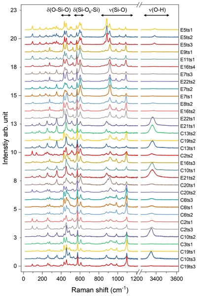
Combined Raman-scattering and wavelength-dispersive electron microprobe (WD-EMP) analyses of natural biotites expanding over the whole biotite solid-solution series demonstrate that the chemical composition of the MO6 octahedra, TO4 tetrahedra, and interlayer space can be non-destructively determined by Raman spectroscopy with relative uncertainties below 8 %. The content of critical minor elements such as Ti at the octahedral site can be quantified as well with a relative error of ~ 20 %.
Combined Raman-scattering and wavelength-dispersive electron microprobe (WD-EMP) analyses of...


