the Creative Commons Attribution 4.0 License.
the Creative Commons Attribution 4.0 License.
 | 19 Jun 2026
Piilonenite-(Nd), NaNd(CO3)2 ⋅ 3H2O, a new neodymium-dominant carbonate mineral from Mont Saint-Hilaire, Quebec, Canada
Inna Lykova
Ralph Rowe
Simon J. Teat
Glenn Poirier
Stephanie Barnes
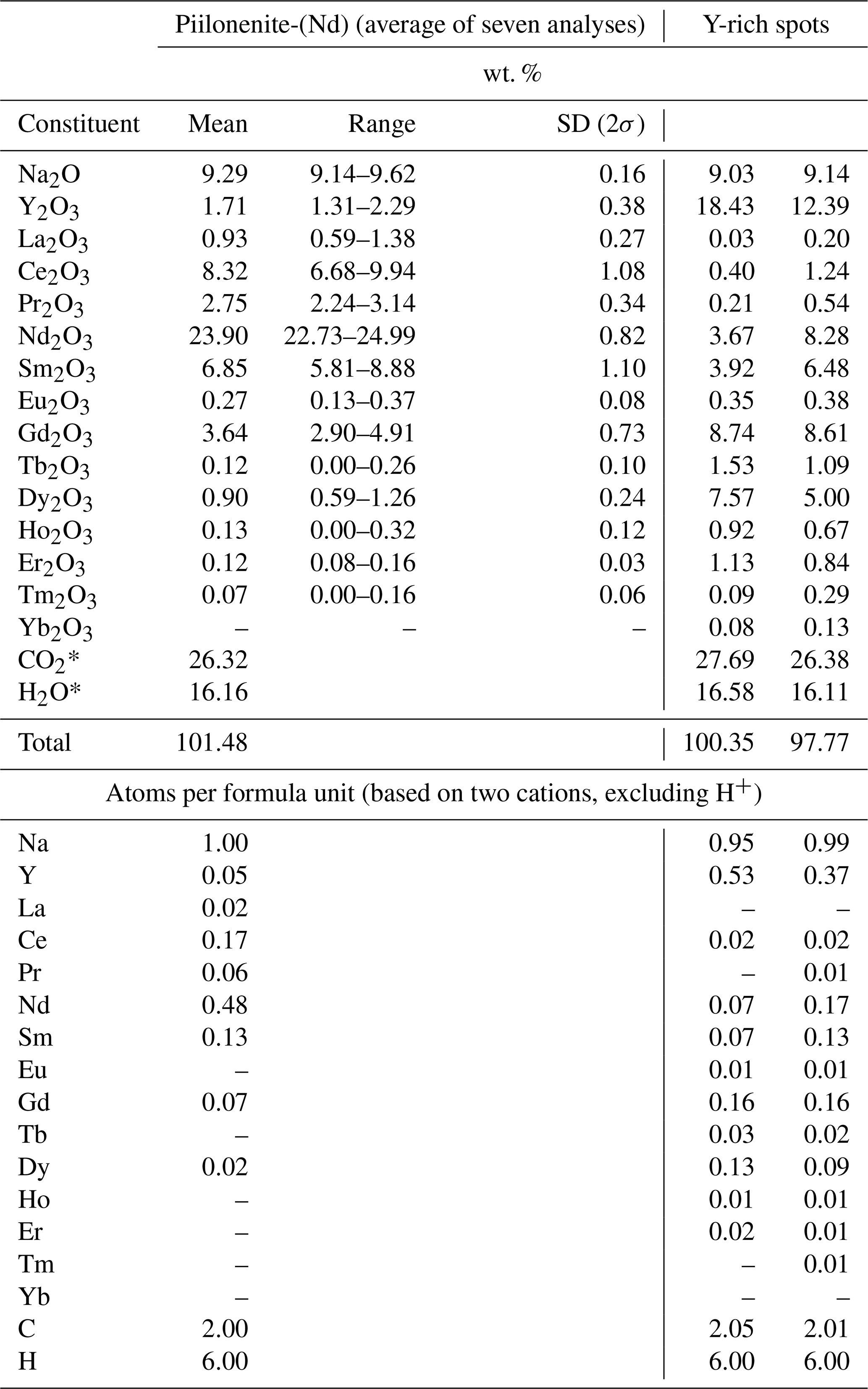
The new mineral piilonenite-(Nd), ideally NaNd(CO3)2⋅3H2O, was found at Mont Saint-Hilaire, Quebec, Canada, in apophyses of the Poudrette pegmatite. It occurs as thin, bladed crystals up to 600 µm in size. Piilonenite-(Nd) is colourless or white. The streak is white; the lustre is vitreous. Cleavage is perfect on {010}. Dcalc is 3.21 g cm−3. Piilonenite-(Nd) is optically biaxial (+), α=1.546(3), β=1.616(3), γ=1.638(3), 2 V (meas.) = 56(3)°, and 2 V (calc.) = 56° (589 nm). The composition (wt. %, average of seven analyses) is Na2O 9.29, Y2O3 1.71, La2O3 0.93, Ce2O3 8.32, Pr2O3 2.75, Nd2O3 23.90, Sm2O3 6.85, Eu2O3 0.27, Gd2O3 3.64, Tb2O3 0.12, Dy2O3 0.90, Ho2O3 0.13, Er2O3 0.12, Tm2O3 0.07, CO2 26.32, H2O 16.16, total 101.48. The empirical formula of the holotype calculated on the basis of two cations is as follows: Na1.00(Nd0.48Ce0.17Sm0.13Gd0.07Pr0.06Y0.05La0.02Dy0.02)∑1.00(CO3)2(H2O)3. The IR spectrum shows IR bands of O–H-stretching and H–O–H bending vibrations of H2O molecules and C–O-stretching vibrations of CO-group molecules. The mineral is orthorhombic, P212121, a=6.7914(11) Å, b=17.135(3) Å, c=6.4360(10) Å, and V=749.0(2) Å3 and Z=4. The strongest reflections of the powder X-ray diffraction pattern [d,Å(I)(hkl)] are as follows: 8.59(100)(020), 4.690(14)(101), 4.291(30)(040), 3.417(10)(200), and 3.175(17)(012, 220, 141). The crystal structure, solved and refined from synchrotron data (R1=0.092), is unique. There are two alternating layers: (1) vertex-sharing Nd-centred polyhedra and “flat-lying” carbonate (CO3)2− groups and (2) chains of vertex-sharing Na-centred polyhedra parallel to (100) and “standing-on-edge” carbonate (CO3)2− groups.
- Article
(5850 KB) - Full-text XML
-
Supplement
(495 KB) - BibTeX
- EndNote


This paper describes piilonenite-(Nd), NaNd(CO3)2⋅3H2O, the second Nd-dominant mineral after bainbridgeite-(NdCe), Na2Ba2NdCe(CO3)6⋅3H2O (Lykova et al., 2023b), found at Mont Saint-Hilaire, Quebec, Canada.
Piilonenite-(Nd) is in named in honour of Paula Piilonen (born 1974), a Canadian mineralogist and research scientist at the Canadian Museum of Nature and a former president of the Mineralogical Association of Canada. Piilonen specializes in the mineralogy of alkaline rocks. She is the senior author of the descriptions of two new mineral species from Mont Saint-Hilaire. The parenthesized suffix (Nd) is added in accordance with the nomenclature for rare earth (yttrium and lanthanides) mineral species established by Levinson (1966).
Both the new mineral and the name have been approved by the Commission on New Minerals, Nomenclature and Classification of the International Mineralogical Association (IMA CNMNC), proposal no. IMA 2025-031. The holotype of piilonenite-(Nd) was deposited in the collection of the Canadian Museum of Nature, Ottawa, Canada. The catalogue number is CMNMC 93393.
Piilonenite-(Nd) was thought to be the unknown phase UK119 from Mont Saint-Hilaire, which was previously investigated by Robert Gault (electron microprobe analyses, 1999), Joel Grice (attempted to solve the structure, 2000), Ralph Rowe (powder X-ray diffraction, 2007), and Igor Pekov (attempted to solve the structure, 2009–2013) (Horváth et al., 2019). However, our examination revealed that “UK119” is, in fact, at least two different phases: one of them is piilonenite-(Nd), and another one is an unrelated Na–Ce carbonate. These were found together at the same time (László Horváth, personal communication, 2025), which led to the confusion. Any information available to date on UK119 could refer to either or both of the minerals.
Piilonenite-(Nd) was found at the Poudrette quarry (Demix quarry) in the famous alkaline agpaitic igneous complex Mont Saint-Hilaire, Quebec, Canada. The material was collected by Elsa Pfenninger-Horváth and László Horváth on 18 August 2000 (László Horváth, personal communication, 2025) in apophyses (3–6 cm wide) of the Poudrette pegmatite cutting the hornfels xenolith on level 8 of the quarry.
The Poudrette pegmatite is by far the largest pegmatite observed in the quarry. It is unusual for the extreme diversity of its minerals and the fact that all of the observed parts of the pegmatite and its extensive apophyses (offshoots) were enclosed in the enormous hornfels xenolith. These vein-like apophyses radiate outward from the main body and can be traced for tens of metres. These contain many of the same minerals as found in the main pegmatite. The mineral assemblages differ in various parts of the pegmatite, varying from silicate- to carbonate-dominant compositions (Horváth et al., 2019, and the references therein).
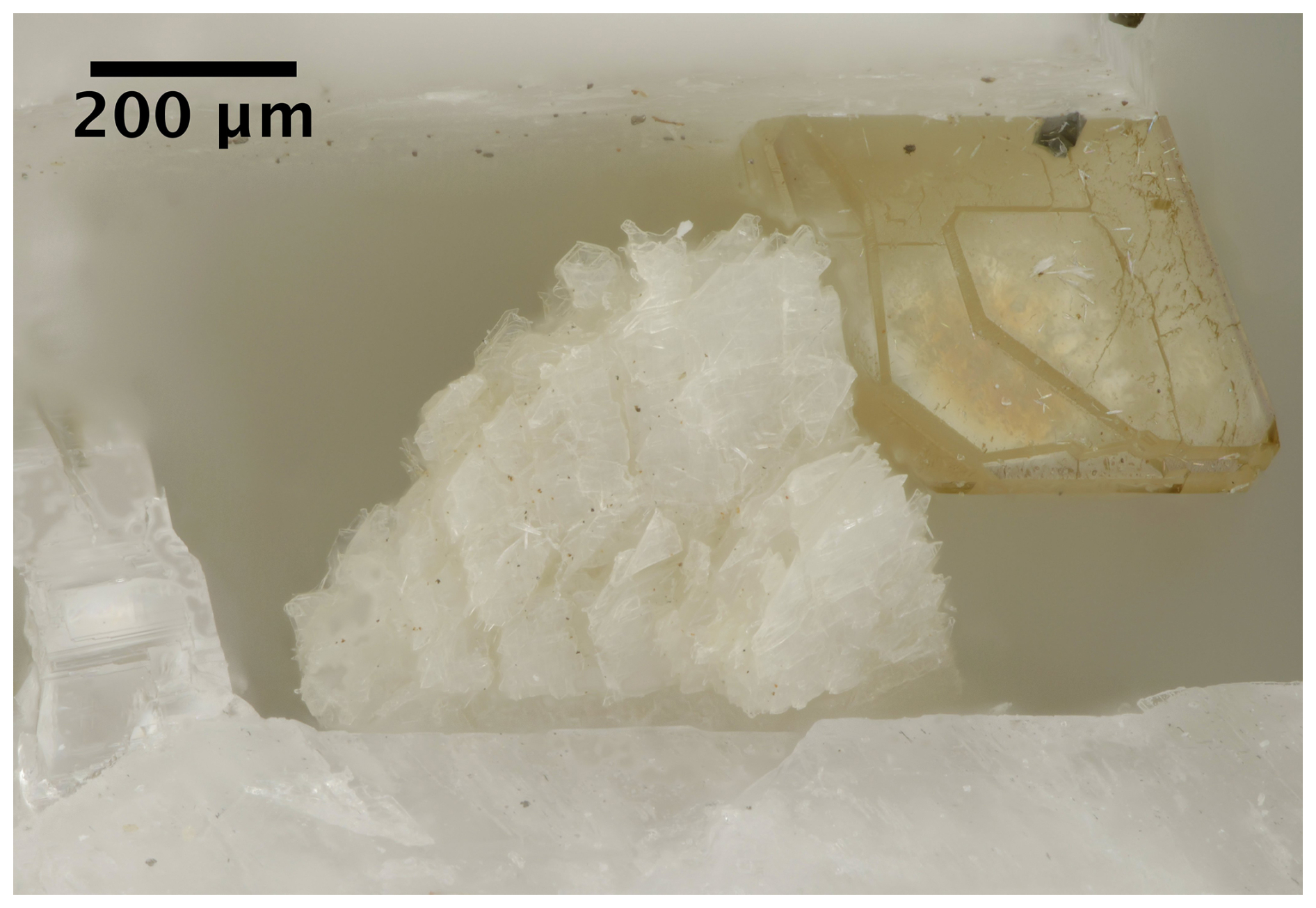
Figure 1White, bladed piilonenite-(Nd) crystal. Specimen CMNMC 93393. Canadian Museum of Nature collection.
Piilonenite-(Nd) occurs as thin, bladed crystals up to 600 µm in size (Fig. 1), together with siderite, calcite, microcline, sphalerite, garronite-Na, quartz, rutile, and aegirine.
Piilonenite-(Nd) is colourless or white (Fig. 1). The streak is white; the lustre is vitreous. The mineral has perfect cleavage on {010}; its fracture is uneven. The Mohs hardness could not be determined as the crystals are very thin and flaky. The mineral is non-fluorescent under ultraviolet light. The density calculated using the empirical formula and unit-cell volume refined from the single-crystal X-ray diffraction (XRD) data is 3.21 g cm−3.
Piilonenite-(Nd) is optically biaxial (+), α=1.546(3), β=1.616(3), γ=1.638(3), 2V (meas.) = 56(3)° (from a spindle-stage extinction curve), and 2 V (calc.) = 56°.
Electron microprobe analyses (EMPA) for piilonenite-(Nd) were obtained using a JEOL 8230 SuperProbe electron microscope equipped with five wavelength dispersive (WDS) spectrometers (University of Ottawa – Canadian Museum of Nature MicroAnalysis Laboratory, Canada) using an accelerating voltage of 20 kV, a beam current of 10 nA, and a beam diameter of 30 µm. Piilonenite-(Nd) is unstable under an electron beam, and so a larger beam diameter was used to minimize element migration. The following reference materials were used: NaInSi2O6 (NaKα), YAG (YLα), LaPO4 (LaLα), CePO4 (CeLα), PrPO4 (PrLβ), NdPO4 (NdLα), SmPO4 (SmLα), EuPO4 (EuLα), GdPO4 (GdLα), TbPO4 (TbLα), DyPO4 (DyLβ), and HoPO4 (HoLβ). The intensity data were corrected for time-dependent intensity (TDI) loss (or gain) using a self-calibrated correction for NaKα, YLα, and LaLα. H2O and CO2 contents were not analysed due to the paucity of the available material.
The Fourier transform infrared (FTIR) spectrum of piilonenite-(Nd) was obtained using a Bruker Hyperion 2000 microscope interfaced to a Tensor 27 spectrometer with a wide-band mercury cadmium telluride (MCT) detector (Canadian Conservation Institute, Canada). A small fragment of piilonenite-(Nd) was mounted on a low-pressure diamond anvil microsample cell and analysed in transmission mode. The spectrum was collected between 4000–400 cm−1, with the co-addition of 256 scans at a 4 cm−1 resolution.
Powder X-ray diffraction (PXRD) data were collected at the Canadian Museum of Nature, Canada, using a Bruker D8 Discover microdiffractometer equipped with a DECTRIS EIGER2 R 500 K detector and I µS microfocus X-ray source ( Å) running at 50 KV and 1000 µA. The detector distance was set at 174.33 mm. The Kα2 contribution was removed using the “Strip Kα2” tool in Bruker Diffrac.EVA V4.3. The instrument was calibrated using a statistical calibration method (Rowe, 2009). A powder ball of piilonenite-(Nd) ∼200 µm in diameter, mounted on a fibre pin mount, was analysed with continuous Phi rotation and 10° rocking motion along the Psi axis of the centric Eulerian cradle stage.
Synchrotron single-crystal X-ray diffraction (SC-XRD) studies were carried out at the Lawrence Berkeley National Laboratory (USA) on Advanced Light Source beamline 12.2.1 with a Bruker D8 with PHOTONII detector using synchrotron radiation (λ=0.7288).
Table 1Chemical data for piilonenite-(Nd).
* Calculated based on the stoichiometry and the structural data, assuming H2O = 3 pfu.
5.1 Chemical data
Chemical data on piilonenite-(Nd) are given in Table 1. The contents of K, Ca, Sr, Ba, Zr, U, Th, Lu, and F are below the detection limit.
The empirical formula calculated on the basis of two cations, excluding H and C, and O = 9 is as follows: Na1.00(Nd0.48Ce0.17Sm0.13Gd0.07Pr0.06Y0.05La0.02Dy0.02)∑1.00
(CO3)2(H2O)3. The simplified formula is Na(Nd,Ce,Sm,Gd,Pr,Y)(CO3)2(H2O)3. The ideal end-member formula is NaNd(CO3)2⋅3H2O, which requires Na2O 9.08, Nd2O3 49.29, CO2 25.79, H2O 15.84, total 100 wt. %.
Although most piilonenite-(Nd) crystals are relatively homogenous, we found several areas with a very high Y content, up to 18.4 wt. % Y2O3 (Table 1), which could correspond to a Y-dominant piilonenite with the simplified formula Na(Y,Gd,Dy,Nd,Sm)(CO3)2(H2O)3. Due to the thinness of the crystals, we were unable to determine spatial relationships between Nd- and Y-dominant areas.
Piilonenite-(Nd) slowly dissolves in an aqueous HCl solution at room temperature with effervescence.
5.2 Infrared spectroscopy
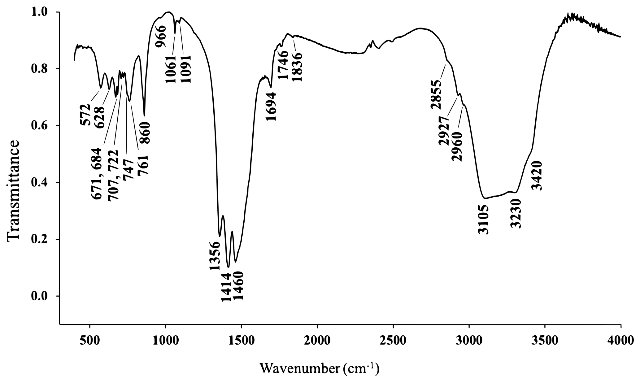
The IR spectrum of piilonenite-(Nd) (Fig. 2) shows IR bands of O–H-stretching (in the range from 3105 to 3230 cm−1) and H–O–H-bending (at 1694 cm−1) vibrations of H2O molecules and C–O-stretching (in the range of 1356–1460 cm−1) vibrations of CO-group molecules. A shoulder at 3420 cm−1 corresponds to O–H-stretching vibrations of basic OH− anions. The bands at 1061 and 1091 cm−1 can be assigned to the non-degenerate mode of C–O-stretching vibrations, indicating polarization of CO groups, as this mode would be inactive if symmetric non-polarized carbonate groups (with a 3-fold axis) were present in the IR spectrum. The band assignment was made in accordance with Chukanov and Chervonnyi (2016).
Table 2X-ray powder diffraction data (d in Å) for piilonenite-(Nd). The strongest reflections are given in bold.
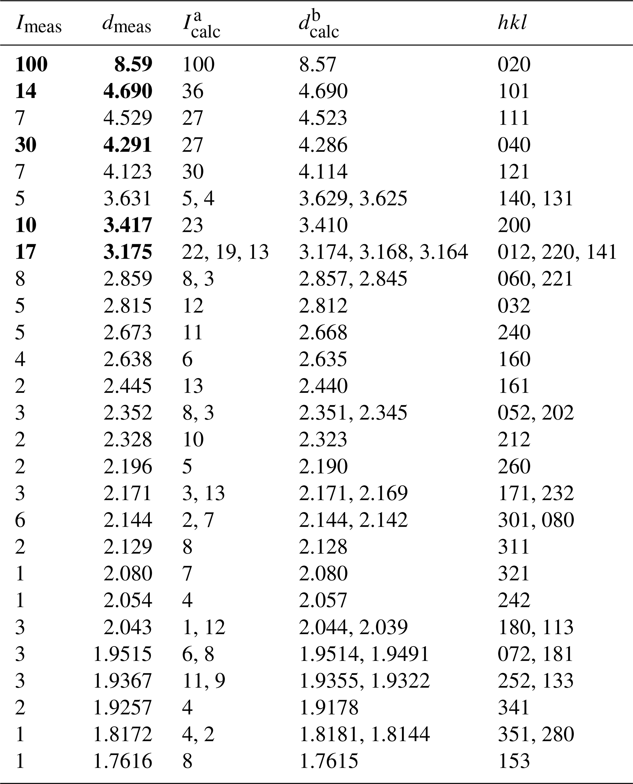
a Calculated from the crystal structure determination. b Calculated from PXRD Rietveld unit-cell refinement with a=6.8201(2), b=17.1439(3), c=6.4590(2) Å, and V=755.21(4) Å3.
5.3 X-ray diffraction data and description of the crystal structure
The indexed PXRD data are given in Table 2. Parameters of the orthorhombic unit cell refined from the data are as follows: a=6.8201(2) Å, b=17.1439(3) Å, c=6.4590(2) Å, and V=755.21(4) Å3. The PXRD pattern in the xy format is available in the Supplement.
Table 3Crystal data, data collection information, and structure refinement details for piilonenite-(Nd).
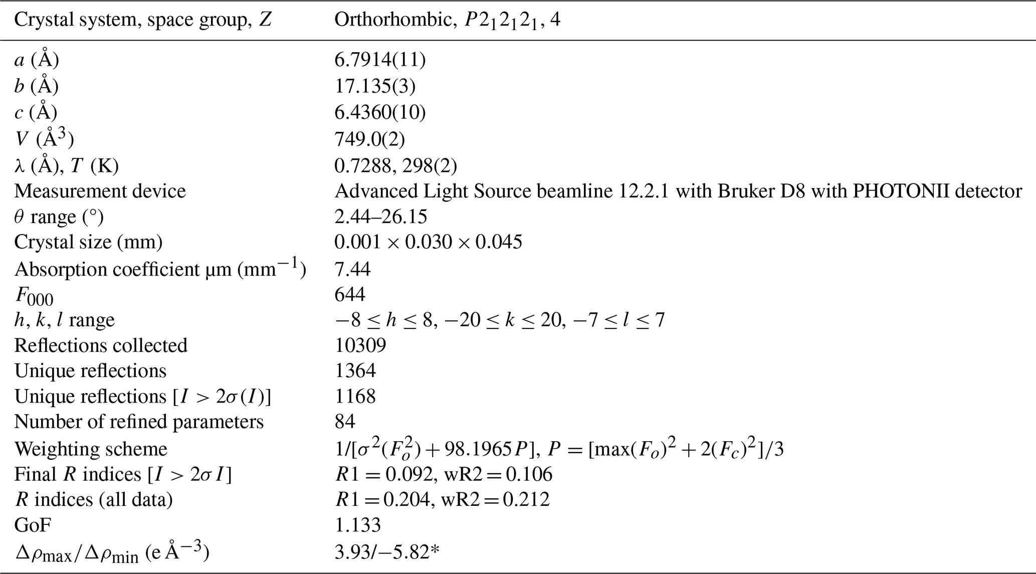
* There are multiple residual electron density peaks and holes near the Nd and C2 sites due to both the crystal quality and data processing issues with heavy elements in a light matrix.
Table 4Coordinates and equivalent displacement parameters (Ueq, in Å2) of atoms and bond valence sums (BVSs) for piilonenite-(Nd).
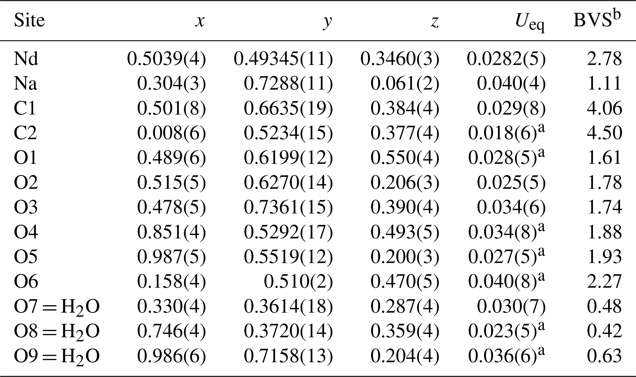
a Uiso. b Bond valence parameters were taken from Gagné and Hawthorne (2015).
Table 5Selected interatomic distances (Å) in the structure of piilonenite-(Nd). The average distances are given in bold.
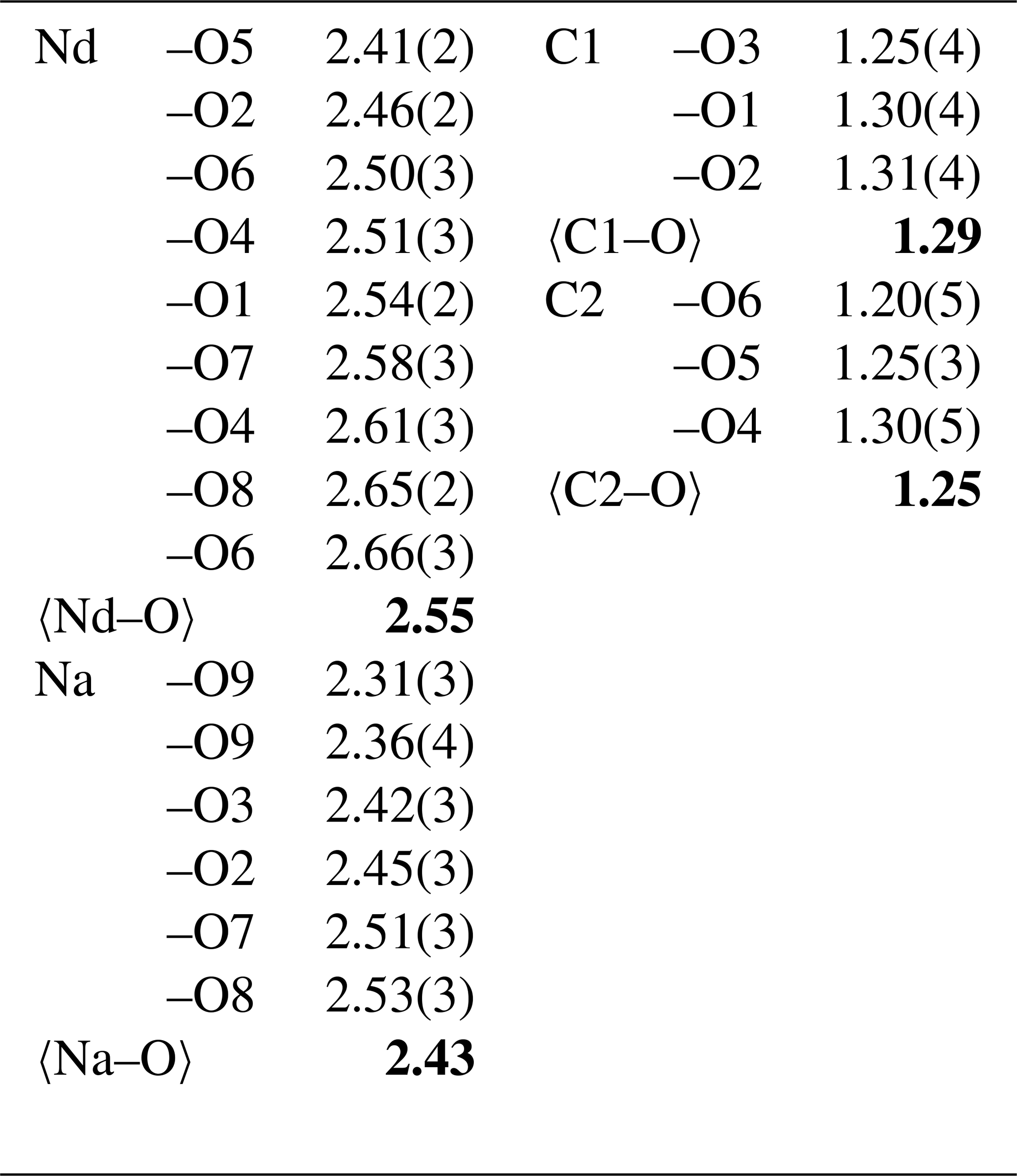
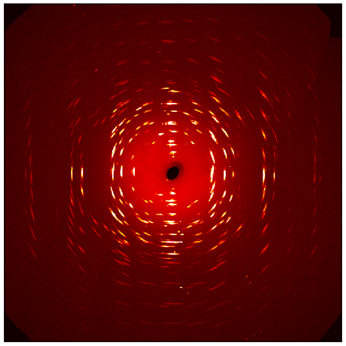
Figure 3Pseudo-procession image in the 0kl reciprocal plane obtained from piilonenite-(Nd) diffraction data.
The single-crystal X-ray diffraction data were indexed in the P212121 space group with the following unit-cell parameters: a=6.7914(11) Å,b = 17.135(3) Å, c=6.4360(10) Å, and V=749.0(2) Å3. The structure was solved and refined (R1=0.092) on the basis of 1364 independent reflections with I>2σ(I) using the SHELXL-2018/3 program package (Sheldrick, 2015). Crystal data, data collection information, and structure refinement details are given in Table 3; atom coordinates, equivalent displacement parameters, and bond valence calculations are given in Table 4; and selected interatomic distances are given in Table 5. The studied crystal demonstrated twinning by merohedry Class I (Nespolo and Ferraris, 2000), an inversion twinning, with the twin domains ratio of 54:46. The crystallographic information file (CIF) for piilonenite-(Nd) is available in the Supplement. It was also deposited in the Inorganic Crystal Structure Database (ICSD; no. CSD 2550589).
Piilonenite-(Nd) crystals exhibit very poor diffraction quality due to significant bending. Two previous attempts to solve its structure by Joel Grice and by Igor Pekov were unsuccessful (Horváth et al., 2019). We were also unable to make any progress with single-crystal diffractometers and attempted to use synchrotron single-crystal X-ray diffraction instead. The obtained data were poor, with multiple split reflections and streaks (Fig. 3). Nevertheless, we were able to solve the structure and proposed a reasonable model that matches well with the PXRD diffraction pattern (Fig. 4).
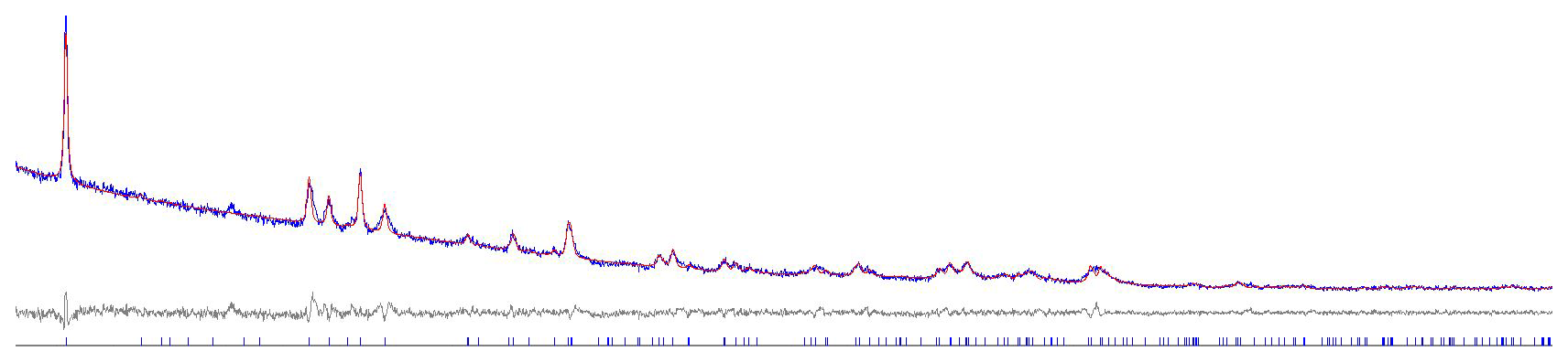
Figure 4Rietveld refinement plot for piilonenite-(Nd) showing the observed (blue), calculated (red), and difference (grey) patterns.
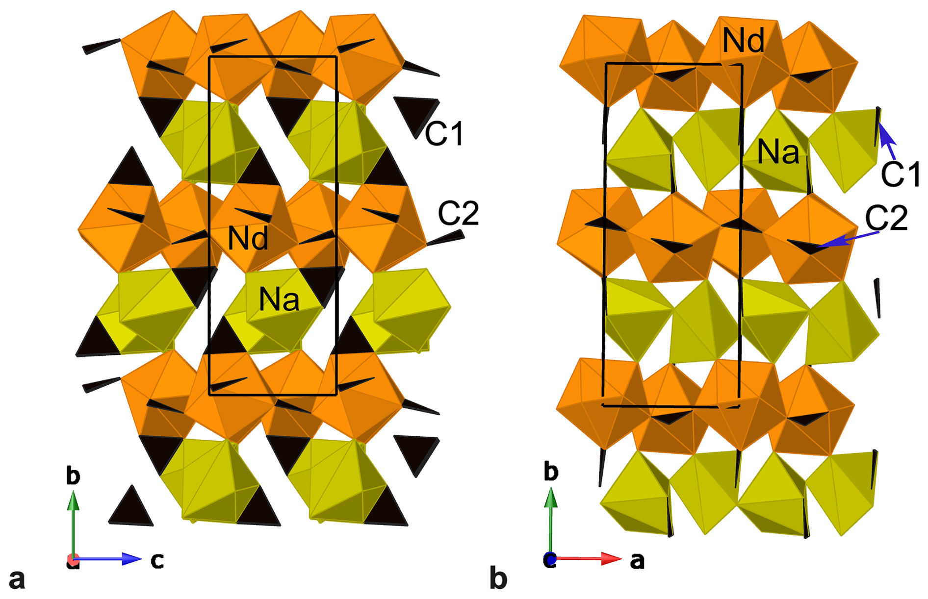
Figure 5General view of the crystal structure of piilonenite-(Nd), projections on (a) (100) and (b) (001). Carbonate groups are black triangles. The unit cells are outlined.
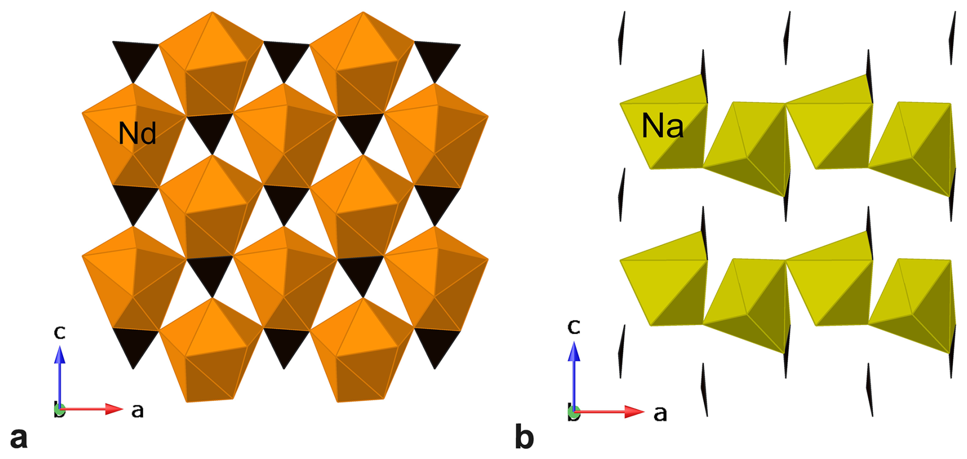
Figure 6A sheet of vertex-sharing Nd-centred polyhedra (a) and chains of vertex-sharing Na-centred octahedra (b) in the structure of piilonenite-(Nd). Carbonate groups are black triangles.
The crystal structure is unique. It is layered on (010) (Fig. 5). There are two alternating layers:
-
a layer of vertex-sharing Nd-centred polyhedra and “flat-lying” carbonate (CO3)2− groups centred by C2 atoms (Fig. 6a),
-
a layer consisting of chains of vertex-sharing Na-centred strongly distorted octahedra parallel to (100) and of “standing-on-edge” carbonate (CO3)2− groups centred by C1 atoms (Figs. 5, 6b).
H2O molecules are present at the O7, O8, and O9 sites (0.48, 0.42, and 0.63 vu, respectively; Table 4). The O9 site connects Na-centred polyhedra into chains, while the O7 and O8 sites connect Nd-centred polyhedra and Na-centred polyhedra in adjacent layers. The estimated contributions of hydrogen bonds based on the O…O distances are given in Table 6. This confirms the presence of H2O molecules at the O7, O8, and O9 sites. This was also confirmed by the presence of the bands of O–H-stretching and H–O–H-bending vibrations in the IR spectrum of piilonenite-(Nd) (Fig. 2).
Table 6Estimated contribution of hydrogen bonds based on the O…O distances for piilonenite-(Nd).
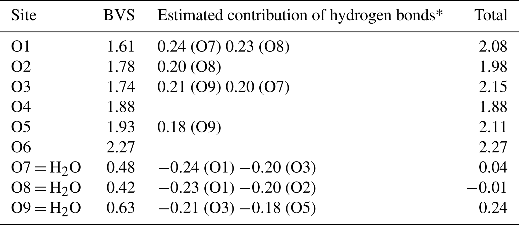
* Based on O…O distances using the function from Ferraris and Ivaldi (1988).
Piilonenite-(Nd) has no closely related minerals or synthetic compounds. There are four water-bearing carbonate minerals, with only sodium and rare earth elements being the dominant cations currently known. Thomasclarkite-(Y), NaY(HCO3)(OH)3⋅4H2O, is a bicarbonate mineral (Grice and Gault, 1998). Shomiokite-(Y), Na3Y(CO3)3⋅3H2O (Khomyakov et al., 1992), and lecoqite-(Y), Na3Y(CO3)3⋅6H2O (Pekov et al., 2010), have a very different stoichiometry (Na : REE = 3:1). Adamsite-(Y), NaY(CO3)2⋅6H2O, has a similar stoichiometry but a higher water content. It also has a layered structure and two types of carbonate groups, namely flat-lying and standing on edge, but the layers have different configurations, with two adjacent Na layers that are connected via hydrogen bonds only, unlike in the structure of piilonenite-(Nd) (Fig. 7; Grice et al., 2000).
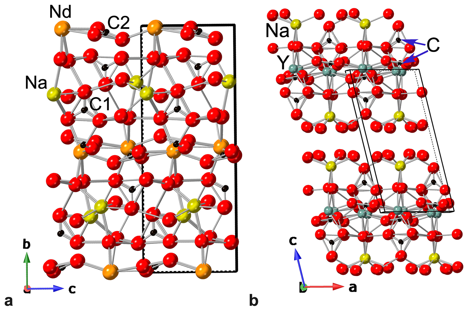
Figure 7Ball-and-stick models of the crystal structures of piilonenite-(Nd) (a) and adamsite-(Y) (b). The unit cells are outlined.
In the vast majority of cases, rare earth elements (REEs; yttrium and lanthanides) are found together in minerals due to their similar chemical properties. This is the basis of the currently used nomenclature for rare earth minerals proposed by Levinson (1966) and later revised by Bayliss and Levinson (1988), which uses a suffix in parentheses to indicate the dominant REE at a site of the mineral structure. Cerium, with over 160 known minerals, and yttrium, with almost 140 minerals, have the largest number of their own minerals. Other REEs usually disperse in them, with larger light REEs going preferentially in Ce-dominant phases, whereas smaller heavy REEs disperse in Y-dominant phases.
Due to its size, Nd3+ occupies an intermediate position between Ce3+ and Y3+ and thus disperses in both Ce- and Y-dominant minerals and rarely forms its own phases. Only 37 Nd-dominant minerals are listed on the official IMA list of minerals (April 2026; IMA-CNMNC, 2026), excluding piilonenite-(Nd).
This is why agpaitic complexes, generally characterized by rich late-stage pegmatite and hydrothermal rare earth mineralization, are not known for diverse Nd minerals, and the few Nd-dominant phases described in such environments are usually products of very specific local conditions.
Rhabdophane-(Nd), Nd(PO4)⋅H2O, and a prospective neodymium analogue of abenakiite-(Ce), Na26Ce6(Si6O18)(PO4)6(CO3)6(SO2)O, were described from the Shkatulka pegmatite, Mt. Alluaiv, Lovozero Massif, Russia (Chukanov et al., 2005). They were found in polymictic pseudomorphs after steenstrupine-(Ce) containing solid bituminous matters and were characterized by both a high content of rare elements and a high degree of their separation in different phases. Carlgieseckeite-(Nd), NaNdCa3(PO4)3F, a belovite-group mineral, and its Ce–Ba-analogue kuannersuite-(Ce), NaCeBa3(PO4)3F0.5Cl0.5, were found in cavities of the same albite vein in the Ilimaussaq complex, Greenland (Pekov et al., 2012). The authors of the description hypothesized that, due to an Sr-poor environment, the Sr-dominant member of the group belovite, NaCeSr3(PO4)3F, the most widespread mineral for this structural type, was not formed, which allowed for the formation of Ca- and Ba-dominant minerals. Ba2+ is significantly larger than Ca2+ and increases the unit-cell volume, which the favoured entry of larger REEs (Ce and La) in kuannersuite, whereas smaller Nd atoms preferentially concentrated in carlgieseckeite. Tundrite-(Nd), Na2Nd2TiO2(SiO4)(CO3)2, was also described from the Ilimaussaq complex, Greenland (Semenov et al., 1967), although the mineral was accepted based on a single chemical analysis without proper investigation and description of the material, and the find has not been replicated yet.
The Nd-dominant phases described above were formed instead of more common Ce-dominant phases of the same structural type when specific local environments allowed for the separation and concentration of Nd.
Piilonenite-(Nd) and two Nd-dominant mckelveyite-group minerals we described recently – bainbridgeite-(NdCe), Na2Ba2NdCe(CO3)6⋅3H2O (IMA2023-018; Lykova et al., 2023b), also from Mont Saint-Hilaire, and mckelveyite-(Nd), NaCaBa3Nd(CO3)6⋅3H2O (IMA2023-017; Lykova et al., 2023a), from the Khibiny Massif, Russia – follow a different pattern. Bainbridgeite-(NdCe) and mckelveyite-(Nd) have more common Y-dominant analogues but not Ce-dominant ones, and there are Y-rich areas (to 0.53 atoms per formula unit of Y) in piilonenite-(Nd) crystals, indicating that the crystal chemically driven affinity of Nd for Y rather than Ce guided the formation of these minerals. In very late-stage hydrothermal processes, with depletion of Y from the solution, Nd can become the prevalent REE in a phase, which resulted in the formation of piilonenite-(Nd); bainbridgeite-(NdCe); mckelveyite-(Nd); and, potentially, other Nd-dominant phases.
Crystallographic data for piilonenite-(Nd) and its PXRD pattern in the xy format are available in the Supplement.
The supplement related to this article is available online at https://doi.org/10.5194/ejm-38-337-2026-supplement.
IL conceptualized the project. RR collected powder X-ray diffraction data. EMPAs were obtained by GP. ST collected synchrotron data. SB obtained the IR spectrum. IL processed the data and interpreted the results. The paper was written by IL with contributions from all of the co-authors.
The contact author has declared that none of the authors has any competing interests.
Publisher's note: Copernicus Publications remains neutral with regard to jurisdictional claims made in the text, published maps, institutional affiliations, or any other geographical representation in this paper. The authors bear the ultimate responsibility for providing appropriate place names. Views expressed in the text are those of the authors and do not necessarily reflect the views of the publisher.
We would like to thank Dan Holtstam and an anonymous referee for their valuable comments and Elsa Pfenninger-Horváth and László Horváth for providing us with the samples for the study. This research used resources of the Advanced Light Source, which is a DOE Office of Science User Facility under contract no. DE-AC02-05CH11231.
This research was financially supported by the Canadian Museum of Nature.
This paper was edited by Sergey Krivovichev and reviewed by Dan Holtstam and one anonymous referee.
Bayliss, P. and Levinson, A. A.: A system of nomenclature for rare-earth mineral species; revision and extension, Am. Mineral., 73, 422–423, 1988.
Chukanov, N. V. and Chervonnyi, A. D.: Infrared Spectroscopy of Minerals and Related Compounds, Springer Cham, Switzerland, 1109 pp., https://doi.org/10.1007/978-3-319-25349-7, 2016.
Chukanov, N. V., Ermolaeva, V. N., Pekov, I. V., Sokolov, S. V., Nekrasov, A. N., and Sokolova, M. N.: Rare-metal mineralization related to bituminous substances in the late pegmatite assemblages of the Khibiny and Lovozero plutons, New Data on Minerals, 40, 80–95, 2005.
Ferraris, G. and Ivaldi, G.: Bond valence vs bond length in O...O hydrogen bonds, Acta Crystallogr. B, 44, 341–344, https://doi.org/10.1107/S0108768188001648, 1988.
Gagné, O. C. and Hawthorne, F. C.: Comprehensive derivation of bond-valence parameters for ion pairs involving oxygen, Acta Crystallogr. B, 71, 562–578, 10.1107/s2052520615016297, 2015.
Grice, J. D. and Gault, R. A.: Thomasclarkite-(Y), a new sodium–rare-earth-element bicarbonate mineral species from Mont Saint-Hilaire, Quebec, Can. Mineral., 36, 1293–1300, 1998.
Grice, J. D., Gault, R. A., Roberts, A. C., and Cooper, M. A.: Adamsite-(Y), a new sodium–yttrium carbonate mineral species from Mont Saint-Hilaire, Quebec, Can. Mineral., 38, 1457–1466, 10.2113/gscanmin.38.6.1457, 2000.
Horváth, L., Gault, R. A., Pfenninger-Horváth, E., and Poirier, G.: Mont Saint-Hilaire: History, Geology, Mineralogy, Can. Mineral. Special Publication 14, Mineralogical Association of Canada, Canada, 634 pp., 2019.
IMA-CNMNC: List of Mineral Names: Updated list of IMA-approved minerals (April 2026), https://cnmnc.units.it/ (last access: 14 April 2026), 2026.
Khomyakov, A. P., Shumyatskaya, N. G., and Polezhaeva, L. T.: Shomiokite-(Y), Na3Y(CO3)3⋅3H2O: a new mineral, Zapiski Vserossijskogo Mineralogicheskogo Obshchestva, 121, 129–132, 1992.
Levinson, A. A.: A system of nomenclature for rare-earth minerals, Am. Mineral., 51, 152–158, 1966.
Lykova, I., Rowe, R., Poirier, G., and Barnes, S.: Mckelveyite-(Nd), IMA 2023-017, in: CNMNC Newsletter 74, Eur. J. Mineral., 35, https://doi.org/10.5194/ejm-35-659-2023, 2023a.
Lykova, I., Rowe, R., Poirier, G., Friis, H., Ojaste, K., and Barnes, S.: Bainbridgeite-(NdCe), IMA 2023-018, in: CNMNC Newsletter 74, Eur. J. Mineral., 35, https://doi.org/10.5194/ejm-35-659-2023, 2023b.
Nespolo, M. and Ferraris, G.: Twinning by syngonic and metric merohedry. Analysis, classification and effects on the diffraction pattern, Zeitschrift für Kristallographie – Crystalline Materials, 215, 77–81, https://doi.org/10.1524/zkri.2000.215.2.77, 2000.
Pekov, I. V., Chukanov, N. V., Zubkova, N. V., Ksenofontov, D. A., Horváth, L. S., Zadov, A. E., and Pushcharovsky, D. Y.: Lecoqite-(Y), Na3Y(CO3)3⋅6H2O, a new mineral species from Mont Saint-Hilaire, Quebec, Canada, Can. Mineral., 48, 95–104, 10.3749/canmin.48.1.95, 2010.
Pekov, I. V., Zubkova, N. V., Husdal, T. A., Kononkova, N. N., Agakhanov, A. A., Zadov, A. E., and Pushcharovsky, D. Y.: Carlgieseckeite-(Nd), NaNdCa3(PO4)3F, a new belovite-group mineral species from the Ilímaussaq alkaline complex, South Greenland, Can. Mineral., 50, 571–580, 10.3749/canmin.50.2.571, 2012.
Rowe, R.: New statistical calibration approach for Bruker AXS D8 Discover microdiffractometer with Hi-Star detector using GADDS software, Powder Diffraction, 24, 263–271, https://doi.org/10.1154/1.3193683, 2009.
Semenov, E. I., Kazakova, M. E., and Aleksandrova, R. A.: 1. The Lovozero minerals – nenadkevichite, gerassimovskite and tundrite - from Ilímaussaq, South Greenland, Meddelelser om Grønland, 181, 1–12, 1967.
Sheldrick, G. M.: Crystal structure refinement with SHELXL, Acta Crystallogr. C, 71, 3–8, 10.1107/s2053229614024218, 2015.