the Creative Commons Attribution 4.0 License.
the Creative Commons Attribution 4.0 License.
 | 07 Oct 2026
Synthesis of nano-porous ellipsoid hematite and the crystal growth mechanism
Meirong Zong

Nshuti Cedrick
Wei Liang
Pinghua Zhu
Dongdong Liang
Haonan Liu
Jiaxin Zou
The crystal growth process directly influences crystal morphology. While the growth process of hematite crystals with specific exposed facets is relatively well understood, the growth mechanism of hematite without distinct facets, particularly porous ellipsoidal hematite, remains unclear. This study systematically investigates the hydrothermal synthesis and crystal growth mechanism of nano-porous ellipsoidal hematite. Under elevated temperatures, iron oxyhydroxide (FeOOH) undergoes a sequential transformation involving dissolution–recrystallization, interfacial nucleation, and solid-state phase transition, ultimately forming hematite. Samples collected at specific time intervals (15, 30, 45, 60, 90, and 120 min) were characterized using X-ray diffraction (XRD), field emission scanning electron microscopy (FE-SEM), transmission electron microscopy (TEM), and high-resolution TEM (HRTEM). The results indicate that lattice rearrangement during the monoclinic-to-hexagonal transition and thermally driven dehydration play critical roles in generating the nano-porous ellipsoidal morphology. This work offers new insights into the controllable hydrothermal synthesis of hematite and its crystallographic transformation pathways.
- Article
(2637 KB) - Full-text XML
- BibTeX
- EndNote


Hematite, also known as ochre, is a common iron ore primarily composed of iron oxide (Fe2O3) (Li et al., 2024; Liu et al., 2024; Wu et al., 2018). Hematite is a common mineral with diverse crystal forms, including flake, spherical, and fibrous structures (Maiti et al., 2015; Liu et al., 2019; Kaufhold et al., 2022). During crystallization, atoms, molecules, or ions are arranged in a specific geometric pattern to form a repeating lattice structure. This structure directly influences the crystal's symmetry and physical properties and plays a decisive role in its chemical purity, shape, and size (Nanev, 2023; Friedrich, 2024). Optimizing synthesis conditions and controlling key factors, such as material morphology and size, play a crucial role in the development of new materials.
The crystal growth process of hematite is influenced by multiple factors, and the growth mechanisms of different crystal facets and morphologies vary. Bandi and Srivastav (2020) first reported the role of stromatolites in the growth of hematite, and Sara Akbari (Akbari et al., 2024) reported the structural interrelationships in this transformation process, discussed in their pioneering work. In addition, Liu et al. (2010) found the microstructure of ferric hydroxide was studied by means of scanning electron microscopy (SEM) and Brunauer–Emmett–Teller (BET), and the characteristics of ferric hydroxide hydrate were measured and evaluated by means of statistical analysis. Zhang et al. (2023) cactualized the microstructure of iron oxide was synthesized by sputtering iron film with oxalic-acid-assisted heat treatment at room temperature. The microstructures such as polyhedrons, globular flowers, leaves, and nano-wires were obtained on the glass substrate at different annealing temperatures (Bandi and Srivastav, 2020). In addition, the possible growth mechanism of transformed hematite nano-particles was also proposed. Soltis et al. (2016) indicate that the formation of iron oxide hematite from the precursor of two-line ferrihydrite requires phase transformation and growth. Seminal studies on iron oxide nucleation and hematite phase transition by Matijević (1979) laid the foundation for understanding FeOOH-to-hematite conversion via precipitation and solid-state transformation. In addition to phase transformation, particle aggregation and oriented attachment mechanisms have been reported to drive the formation of porous or anisotropic morphologies in oxide systems such as SiO2 and Fe2O3. These processes facilitate the alignment of primary nano-particles into mesocrystals, followed by lattice fusion under hydrothermal stress, as described in prior works (Sugimoto and Muramatsu, 1996). However, these classical works primarily investigated non-porous morphologies under ambient or low-pressure conditions. Our study extends this scope by focusing on nano-porous ellipsoidal hematite formed under hydrothermal constraints, wherein both interfacial nucleation and anisotropic lattice rearrangement drive pore formation.
Although previous studies have explored the formation pathways of hematite nano-structures, the crystallographic transformation from acicular iron oxyhydroxide to nano-porous hematite remains poorly understood. In particular, the structural rearrangement during the monoclinic-to-hexagonal transition and its role in pore formation have not been clearly elucidated. This study aims to bridge that gap by investigating the morphological and structural evolution of FeOOH under hydrothermal conditions. We focus on identifying the specific intermediate phases and phase transitions, as well as understanding how solid-state transformation governs the final porous morphology of hematite particles. This study prepared nano-porous spherical hematite through hydrothermal synthesis and observed the morphology of the samples at different times. A large amount of needle-shaped iron hydroxide is generated in the early stage of the reaction. As the heating time increases, iron hydroxide undergoes dehydration reaction, and water molecules are removed from the iron hydroxide. At the same time, hydroxylated iron oxide aggregates into ellipsoids. With increasing heating time, hematite ellipsoids are formed through solid-phase transformation and dissolution recrystallization. Some compounds or volatile substances in the hematite ellipsoidal structure are released, ultimately forming nano-porous spherical hematite. Through these results, we explored the growth mechanism of iron hydroxide in hematite, providing a theoretical basis for subsequent research on mineral crystal growth.
2.1 Chemicals and materials
All chemicals are of analytical-grade purity, suitable for direct use without any treatment: ferric chloride hexahydrate (FeCl3-6H2O analytically pure, Guangzhou Jinyuan Chemical Co., LTD.), anhydrous ethanol (C2H6O, analytically pure, Sinopod Chemical Reagents Co., LTD.), methylene blue (C16H18CIN3S-3H2O, analytically pure, Tianjin Dengfeng Chemical Reagent Factory), sodium hydroxide (NaOH, analytically pure, Sinopod Chemical Reagent Co., LTD.), and deionized water.
2.2 Synthesis of nano-ellipsoidal hematite
Nano-sized hematite was synthesized using a hydrothermal method. The preparation of the ethanol solution was as follows: firstly, we measured 144 mL of ethanol solution, added 6 mL of deionized water solution, prepared 150 mL of 96 % ethanol solution placed in the reaction flask, sealed with cling film, and stored temporarily to prevent the liquid from evaporating. We weighed FeCl3-6H2O 4.05 g, put this into 150 mL 96 % ethanol solution, stirred with a magnetic stirrer until completely dissolved, and prepared as 0.1 mol L−1 FeCl3–6H2O solution. The solution was continuously stirred at 300 rpm using a magnetic stirrer before hydrothermal treatment. We then sequentially extracted 12 mL of ferric chloride solution and placed this in a high-pressure reactor (volume: 25 mL) lined with PTFE and then sealed and placed this in the 180 °C oven reaction for 6 h. The rate of temperature rise was 5 °C min−1. The reaction was completed and cooled to room temperature, and then we poured off the supernatant and added deionized water for cleaning, followed by suspension through high-speed separation before being centrifuged and cleaned with deionized water and then centrifuged again to pour off the supernatant. The above steps were repeated 10 times before the solution was put into the oven and dried at 35 °C to obtain nano-pore ellipsoidal spherical hematite powder.
2.3 Characterization of hematite phase, morphology, and transformation
2.3.1 X-ray diffraction
X-ray diffraction (XRD) analyses were carried out by the press-flake method, and the test equipment was an X-ray powder diffractometer (D/MAX2500, Japan). The test parameters were a Cu rotary target (λ=1.5406 Å) with a maximum power of 18 KW and a 2θ angle test range from ∼0–80°, and XRD spectra were collected by continuous step scanning with a step size of 0.02° and a time of 30 s. The XRD spectra were analysed by the X-ray powder diffractometer (D/MAX2500, Japan). The software for physical-phase analysis was JADE 9.5.1. The XRD spectra were fitted using a pseudo-Voigt function for microstructural analysis.
2.3.2 Scanning electron microscopy
The morphology and size of the particles were observed using a field emission scanning electron microscope FE-SEM (Zeiss SUPRA-55, Germany), with a resolution of 0.8 nm, 15 kV and 1.6 nm, 1 kV; an accelerating voltage of 0.02–30 kV, magnification of 12–1 000 000×, and a probe current of 4 pA–20 nA. The Nano Measurer 1.2 was used to calculate the size of each particle. A total of no fewer than 50 particles were analysed using the Nano Measurer 1.2 software for statistical accuracy. The SEM samples were prepared by dropping a suspension of particles prepared by dispersion in water under ultrasonic conditions onto a clean monocrystalline silicon plate, which was then blown dry with a blow-dust ball; the plate was cleaned by ultrasonic cleaning in water and then in ethanol for 10 min before being cleaned with ultrapure water and stored in a sealed container.
2.3.3 Scanning/transmission electron microscopy
A transmission electron microscope (TEM, JEOL JEM-2100, Japan) was used for high-resolution microscopy. The samples were then sprayed with gold to limit charge interference during imaging. Samples for TEM observation were prepared by drop-casting the suspension onto a TEM copper grid with a lace carbon film (300 mesh, Ted Pella, Inc).
2.3.4 Measure sample size
The Nano Measurer 1.2 software was used to measure the size of nano-porous ellipsoid hematite and the surface pore in hematite particles. The measurement method of the Nano Measurer 1.2 software is based on the scale of SEM images. We then obtained the length of the marked dimension line of the test target and calculated the average value of all the data further, and these average data were the final dimension obtained. The size of the nano-porous ellipsoid hematite was obtained by 100 particles. The distribution of the average nano-porous size on the surface of hematite was obtained by 200 pore points from 20 hematite particles.
3.1 Phase evolution revealed by XRD
The XRD pattern of experimental products at 15 and 30 min shows characteristic peaks of FeOOH(ICSD: 31136), including reflections at 2θ=21.2, 33.3, and 36.6°, corresponding to the (110), (021), and (111) planes, respectively. As the reaction progresses to 45 and 60 min, these peaks gradually decrease, while the characteristic peaks of α-Fe2O3 begin to emerge at 2θ=24.1, 33.2, 35.6, 49.5, and 54.1°, indicating phase transformation. At 90 and 120 min, the diffraction peaks of FeOOH disappear entirely, confirming its complete transformation into hematite. This evolution supports a dissolution–recrystallization pathway followed by a solid-state transformation. As ferric chloride reacts with water to form different iron oxides or iron hydroxides, ferric hydroxide precipitates as a colloidal substance, which can cause the solution to become cloudy in the initial stage. As the temperature and heating time increase, ethanol reacts with iron hydroxide to form an ethanol complex. Finally, the precipitate undergoes a hydrothermal reaction at high temperature to obtain pure-phase hematite. This is confirmed by XRD analysis (Fig. 1). After heating for about 30 min, FeOOH is first formed in the solution. The obtained XRD spectra are in good agreement with the standard hematite XRD spectra (ICSD: 98-041-5251). This transformation process clearly demonstrates the sequential phase evolution from FeOOH to α-Fe2O3, driven by temperature-induced dehydration and structural rearrangement.
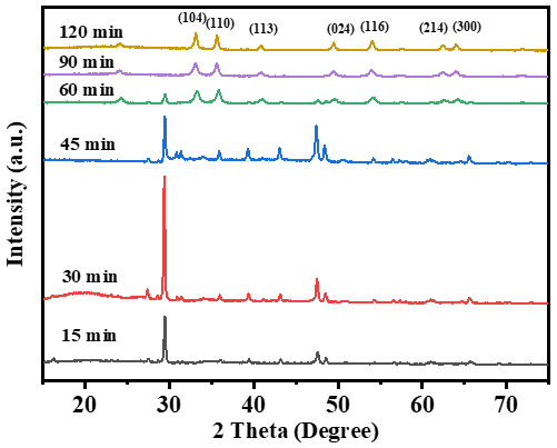
Figure 1XRD patterns of products at different ageing times. The initial product is ferric hydroxide and the final product is hematite.
3.2 Morphological evolution at different times
In the early stage of the reaction, the results indicate that at the beginning of the reaction, a large amount of needle-shaped FeOOH with an average grain size 43 nm formed (Fig. 2a, b), which is consistent with previously reported (Liu et al., 2008). As the reaction time increases, FeOOH forms aggregates (Figs. 2c and 4a) and hematite nucleates (Fig. 4b) and grows on the surface of the aggregates (Fig. 2c). After continuous heating at high temperatures, the iron hydroxide aggregates grew into 0.68±0.1 µm elliptical hematite particles with a width of 0.42±0.1 µm (Figs. 2d to f and 3a). The crystal structure changes under high-temperature heating, and the interior of the crystal contains some gas or volatile substances. These gases may be released under high-temperature conditions, causing pores to form on the crystal surface. Finally, nano-pore ellipsoidal hematite grew (Fig. 2f). The hematite particles have a surface pore diameter of 27.36±3 nm (Fig. 3b). This is very consistent with the probability proposed by Das and Hendry (2014), Cha et al. (2011), and Khani and Ebrahim (2023).
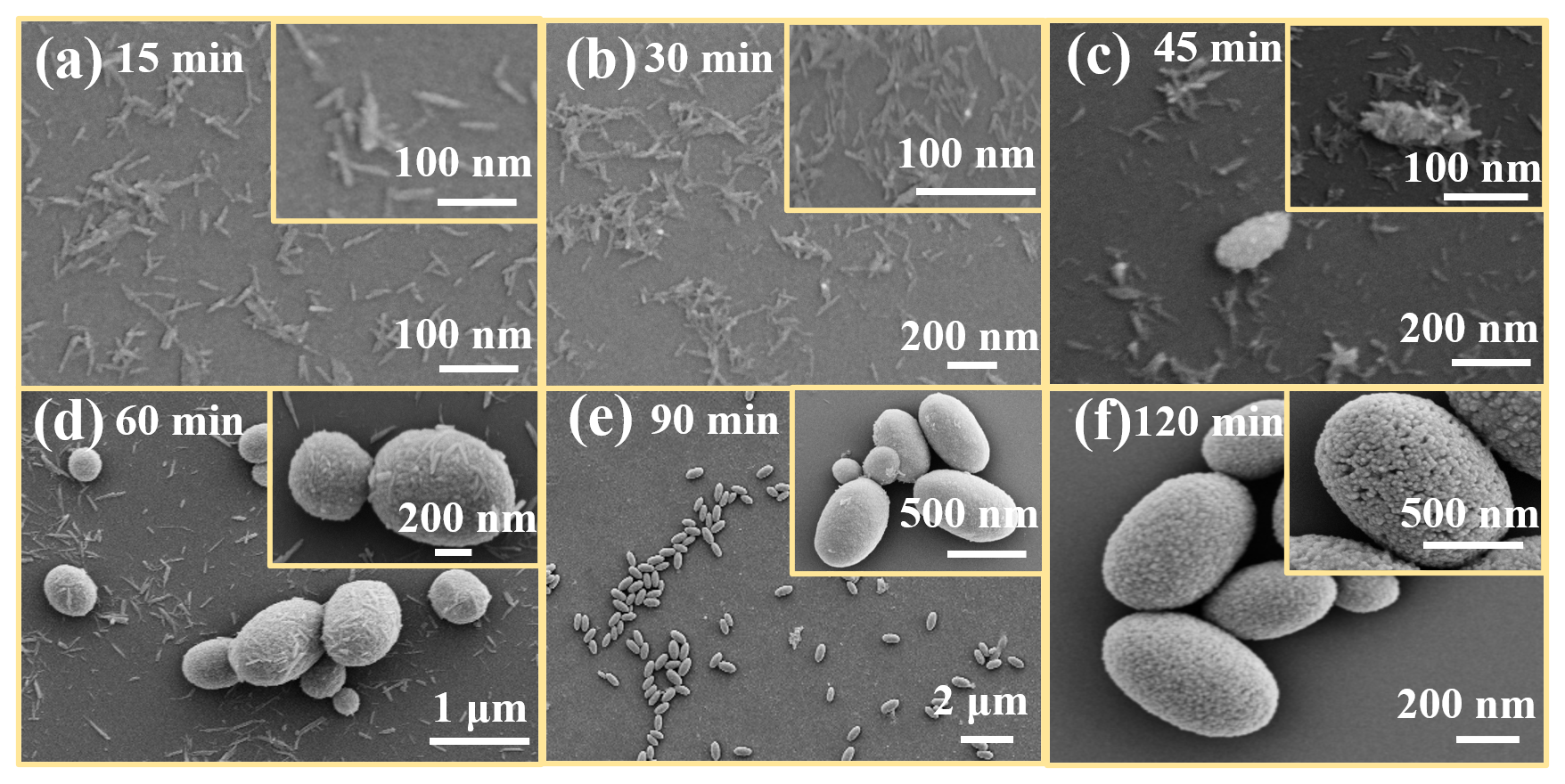
Figure 2Field emission SEM images of samples collected at different hydrothermal reaction times: (a) 15 min – nucleation of short rod-like ferric hydroxide; (b) 30 min – development of dense acicular α-FeOOH structures; (c) 45 min – partial dissolution of FeOOH and formation of aggregated clusters; (d) 60 min – emergence of initial ellipsoidal particles; (e) 90 min – further growth with increasing pore definition; (f) 120 min – fully developed nano-porous ellipsoidal hematite with roughened surfaces and visible porosity.
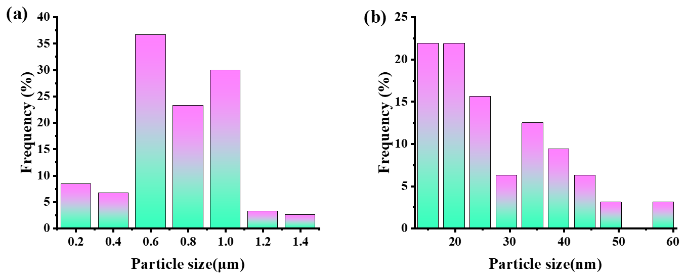
Figure 3(a) Particle size distribution histogram of hematite particles synthesized at 120 min, showing an average length of 0.68±0.1 µm and a width of 0.42±0.1 µm. (b) Pore size distribution derived from nitrogen adsorption data, indicating dominant pore diameters around 27.36±3 nm.
Based on Figs. 2c and 4b, it can be observed that FeOOH aggregates are formed first, and hematite nucleates and grows on the aggregates. The initial aggregates consist of two phases: FeOOH and hematite. As the reaction time increases, the FeOOH content in the aggregates undergoes solid-phase transformation into hematite (Fig. 4c, d), and the free FeOOH in the solution dissolves, promoting the growth of hematite. The conversion of iron hydroxide into hematite involves simultaneous transformation and growth, forming highly crystalline nano-porous ellipsoidal hematite particles (Jiang et al., 2016; Liu et al., 2005; Notini et al., 2022; Lamberov et al., 2012; Ha et al., 2014). The transition of ferric hydroxide into hematite is considered to be a dehydroxy-induced structural transition, and the surface area of the particles increases with this transition (Jian-ming et al., 2009; Sugimoto and Muramatsu, 1996). During the heating process, parts of the surface of the hematite crystals dissolve and then recrystallize in the solution to form a new structure, resulting in the formation of pores on the surface of the hematite (Cha et al., 2011; Khani and Ebrahim, 2023).
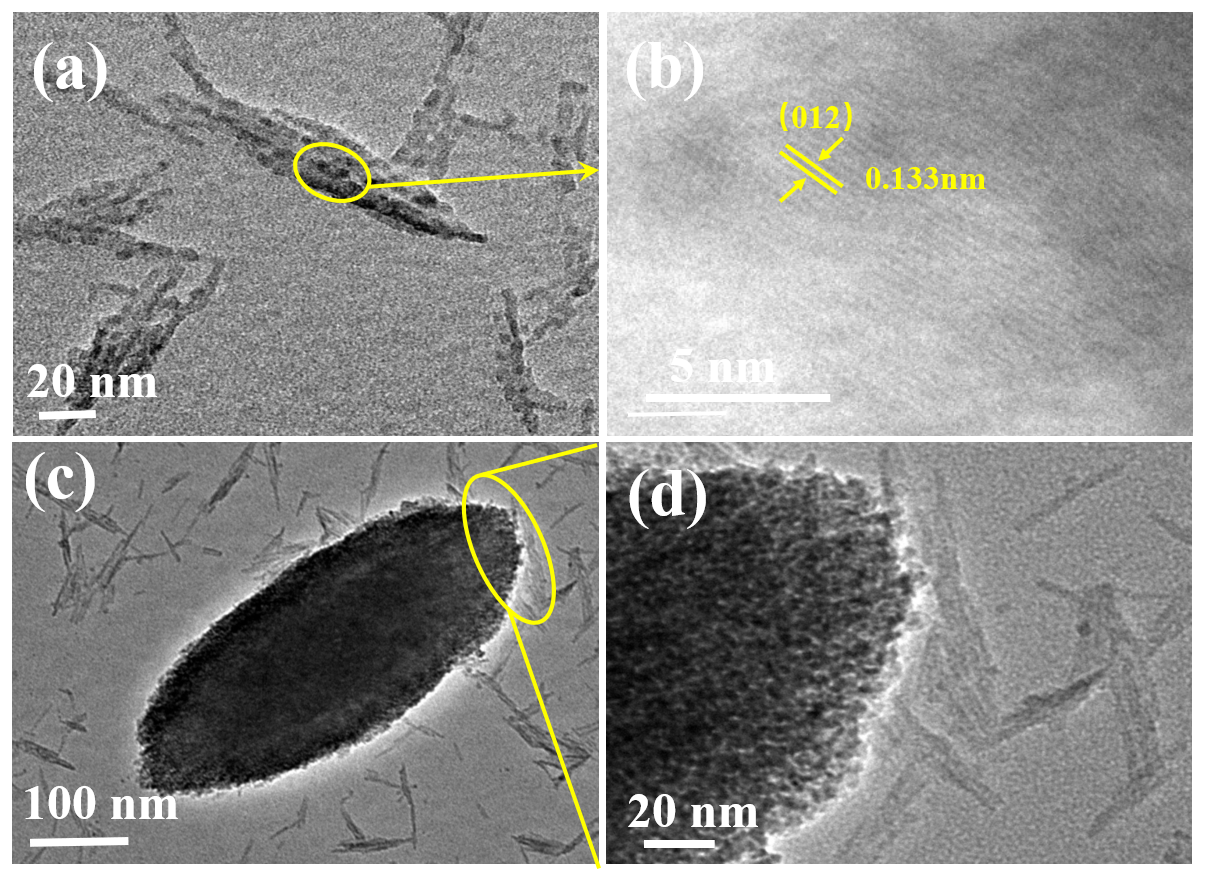
Figure 4Panels (a) and (b) show the 30 min sample; the processes of iron hydroxide dissolution and recrystallization form new nucleation sites. The 45 min sample is shown in panels (c) and (d); it represents the nucleation process where solid-phase transformation and dissolution–recrystallization coexist on the surface of hematite.
3.3 Crystal growth mechanism
For the growth mechanism of nano-porous ellipsoids, it is first necessary to clarify the formation of FeOOH aggregates and the stage of phase transition. The rice-like nano-porous morphology may arise from oriented attachment and aggregation-induced self-assembly mechanisms widely reported in oxide systems (Iler, 1979; Hamada and Matijević, 1981; Sugimoto and Muramatsu, 1996).Since smaller ferric hydroxide does not have ordered vacancies, fewer particles may be formed through interfacial nucleation (Pan et al., 2020). So the formation of FeOOH aggregates laid the foundation for the final morphology of ellipsoidal hematite. According to the XRD pattern and SEM results in this article, phase transition and aggregation occur simultaneously. As iron hydroxide is a metastable phase, it is more prone to phase transition than hematite at higher temperatures and surface energies. However, the initial aggregates are still dominated by FeOOH. Mendili et al. (2012) also confirmed that the crystal lattice parameters of ferric hydroxide hydrate may change under high-temperature heating, and sufficient energy can be provided by increasing the temperature to dissociate the hydrate and release water molecules. These processes facilitate the alignment of primary nano-particles into mesocrystals, followed by lattice fusion under hydrothermal stress. According to Gibbs free-energy data (Cornell and Schwertmann, 2003), hematite becomes the thermodynamically favoured phase beyond ∼80 °C, thereby corroborating our observation of complete FeOOH-to-α-Fe2O3 conversion after 120 min of hydrothermal ageing.
Therefore, we need to explain the form mechanism of the porous structure on hematite particles. Vu et al. (2014) emphasized that the repacking of the oxygen atomic layer from the unidimensional to the hexagonal (hexagonal closest packing) is inevitable during the transition of iron hydroxide into hematite. The rhombohedral crystal structure of FeOOH showed that iron (Fe) forms an octahedral structure with oxygen (O) and hydrogen (H) (Fig. 5a) in hematite (Fig. 5b), in which each Fe atom forms an ionic bond with the surrounding oxygen atom (Soboleva et al., 2022; Supattarasakda et al., 2013). In the transition from a rhombohedral into a hexagonal system, the atoms need to be rearranged to fit the new crystal structure (Shi and Xu, 2014; Meijer and Rossi, 2021). This involves moving atoms from a rhombohedral system to a tightly packed hexagonal energy level in a hexagonal structure. During this transition, dehydration leads to the collapse of hydrogen-bonded water layers and reconfigurations of Fe–O coordination (Hayes et al., 2016). The removal of structural water increases internal stress, resulting in anisotropic lattice contraction or distortion. These local distortions create structural mismatches and voids, which subsequently manifest as surface and internal pores. Therefore, pore formation in nano-ellipsoidal hematite is not merely a morphological byproduct but is inherently coupled with the crystallographic transition from monoclinic into hexagonal symmetry.
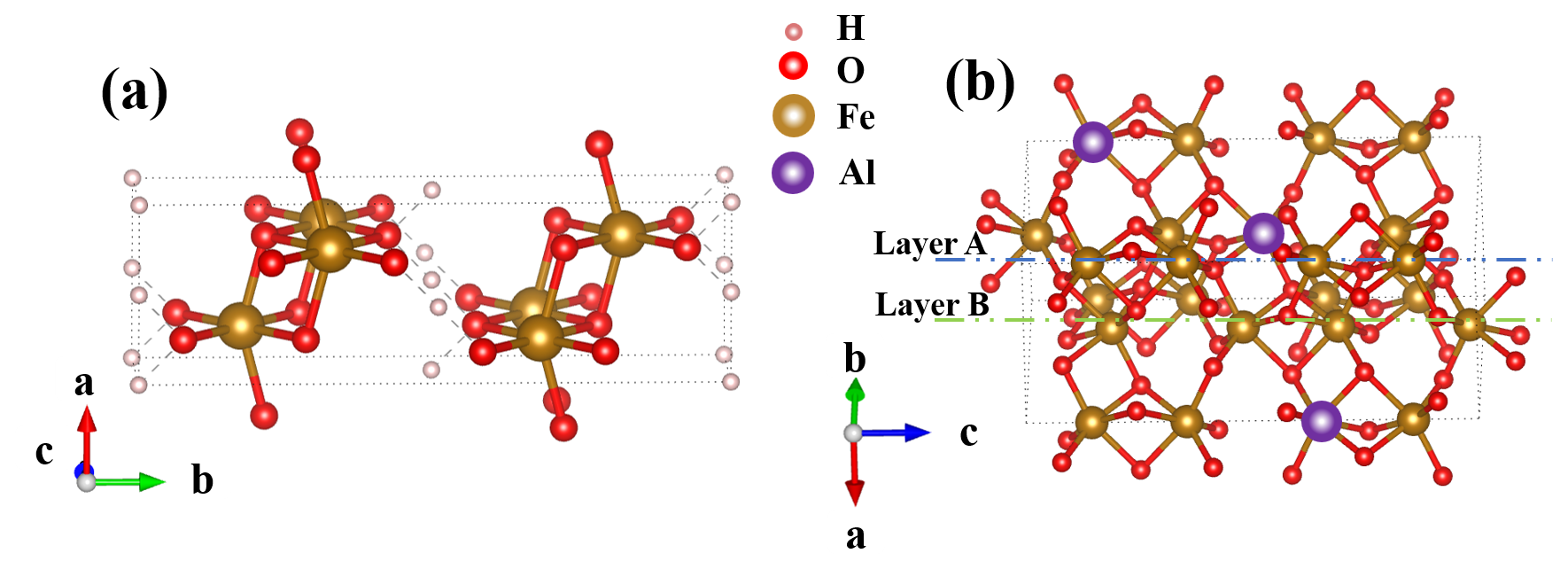
Figure 5(a) Crystal structure diagram of FeOOH and (b) crystal structure diagram of hematite.
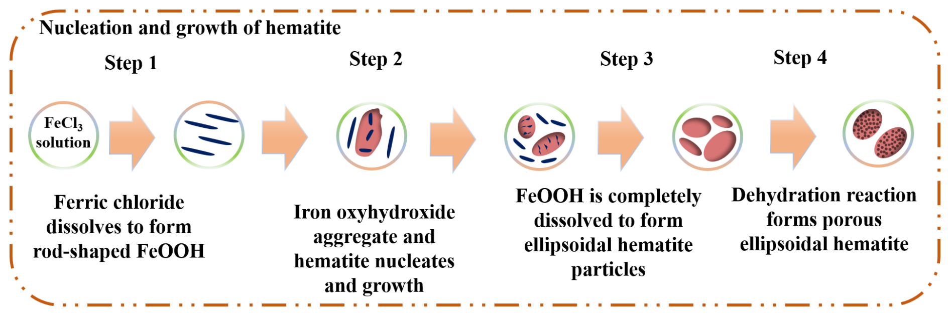
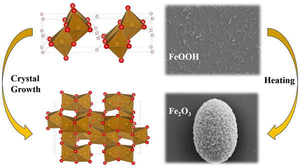
In summary, as shown in Fig. 6, the crystal growth mechanism of nano-porous elliptical hematite can be divided into four stages: (1) Fe3+ nucleates and grows in solution to form needle-shaped FeOOH. (2) FeOOH crystals grow and aggregate into a micro-ellipsoid structure. (3) Under the combined action of solid-phase transformation and dissolution recrystallization, FeOOH is transformed into hematite. (4) Under thermal stress, hematite particles undergo dehydration and lattice rearrangement to drive the formation of pore structures, ultimately transforming into ellipsoids containing nano-pores.
This study elucidates the hydrothermal formation mechanism and crystal growth pathway of nano-porous ellipsoidal hematite derived from FeOOH precursors. It was demonstrated that the transformation proceeds via a multi-step process involving dissolution–recrystallization, interfacial nucleation, and solid-state phase transition. Structural analysis confirmed that the monoclinic-to-hexagonal transformation is accompanied by lattice rearrangement and dehydration, which jointly contribute to the generation of nano-pores on the particle surface. These findings not only provide new insights into the crystallographic evolution of iron oxide minerals but also offer a potential route for the controlled synthesis of porous hematite with specific morphologies and properties. This work holds significant implications for mineralogy, geochemistry, and advanced material fabrication.
No data sets were used in this article.
MZ and WL contributed to the conceptualization of this study. HL and PZ were responsible for data processing. WL and DL performed chemical synthesis and data analysis. WL and MZ wrote the first draft of the paper. NC and MZ were responsible for revising the paper. JZ and PZ participated in drawing the figures and tables.
The contact author has declared that none of the authors has any competing interests.
Publisher's note: Copernicus Publications remains neutral with regard to jurisdictional claims made in the text, published maps, institutional affiliations, or any other geographical representation in this paper. The authors bear the ultimate responsibility for providing appropriate place names. Views expressed in the text are those of the authors and do not necessarily reflect the views of the publisher.
This project was supported by the National Natural Science Foundation of China Youth Fund (grant no. 42202034) and the Open Fund of the Key Laboratory of Solid Waste Treatment and Resource Utilization of the Ministry of Education (Southwest University of Science and Technology) (grant no. 22kfgk03).
This research has been supported by the National Natural Science Foundation of China (NSFC) (grant no. 42202034).
This paper was edited by Elena Belluso and reviewed by lei gao and two anonymous referees.
Akbari, S., Zabihollahi, S., Yaqoubnejad, P., Palandi, Z. K., and Taghavijeloudar, M.: Insight into the roles of hematite iron oxide nanoparticles on microalgae growth, urban wastewater treatment and bioproducts generation: Gompertz simulation, nutrient mass balance and gene expression, Bioresource Technol., 394, 130300, https://doi.org/10.1016/j.biortech.2024.130300, 2024.
Bandi, S. and Srivastav, A. K.: Understanding the growth mechanism of hematite nanoparticles: The role of maghemite as an intermediate phase, Cryst. Growth Des., 21, 16–22, https://doi.org/10.1021/acs.cgd.0c01226, 2020.
Cha, H. G., Kim, S. J., Lee, K. J., Jung, M. H., and Kang, Y. S.: Single-crystalline porous hematite nanorods: photocatalytic and magnetic properties, The Journal of Physical Chemistry C, 115, 19129–19135, https://doi.org/10.1021/jp206958g, 2011.
Cornell, R. M. and Schwertmann, U.: The iron oxides: structure, properties, reactions, occurrences, and uses, vol. 664, Wiley-vch, Weinheim, https://doi.org/10.1515/CORRREV.1997.15.3-4.533, 2003.
Das, S. and Hendry, M. J.: Characterization of hematite nanoparticles synthesized via two different pathways, J. Nanopart. Res., 16, https://doi.org/10.1007/s11051-014-2535-7, 2014.
Friedrich, J.: Methods for bulk growth of inorganic crystals, Encyclopedia of Condensed Matter Physics, 5, 190–207, https://doi.org/10.1016/B978-0-323-90800-9.00087-1, 2024.
Ha, D. H., Caldwell, A. H., Ward, M. J., Honrao, S., Mathew, K., Hovden, R., Koker, M. K. A., Muller, D. A., Hennig, R. G., and Robinson, R. D.: Solid-solid phase transformations induced through cation exchange and strain in 2D heterostructured copper sulfide nanocrystals, Nano Lett., 14, 7090–7099, https://doi.org/10.1021/acs.est.2c0392510.1021/nl5035607, 2014.
Hamada, S. and Matijevć, E.: Ferric hydrous oxide sols. IV. Preparation of uniform cubic hematite particles by hydrolysis of ferric chloride in alcohol–water solutions, J. Colloid Interf. Sci., 84, 274–277, https://doi.org/10.1016/0021-9797(81)90287-3, 1981.
Hayes, D., Hadt, R. G., Emery, J. D., Cordones, A. A., Martinson, A. B. F., Shelby, M. L., Fransted, K. A., Dahlberg, P. D., Hong, J., Zhang, X., Kong, Q., Schoenlein, R. W., and Chen, L. X.: Electronic and nuclear contributions to time-resolved optical and X-ray absorption spectra of hematite and insights into photoelectrochemical performance, Energ. Environ. Sci., 9, 3754–3769, https://doi.org/10.1039/c6ee02266a, 2016.
Iler, R. K.: The Chemistry of Silica: Solubility, Polymerization, Colloid and Surface Properties and Biochemistry of Silica, colloid & surface properties & biochemistry, Wiley-Interscience, New York, 1979.
Jiang, Z., Liu, Q., Dekkers, M. J., Barrón, V., Torrent, J., and Roberts, A. P.: Control of earth-like magnetic fields on the transformation of ferrihydrite to hematite and goethite, Sci. Rep., 6, 30395, https://doi.org/10.1038/srep30395, 2016.
Jian-ming, P., Pei-min, G., Pei, Z., Chao-zhen, C., and Dian-wei, Z.: Influence of size of hematite powder on its reduction kinetics by H2 at low temperature, Iron and Steel Research Journal, 16, 7–11, https://doi.org/10.1016/s1006-706x(10)60002-7, 2009.
Kaufhold, S., Ufer, K., Hein, M., Götze, N., and Dohrmann, R.: A combined IR and XRD study of natural well crystalline goethites (α-FeOOH), Acta Geochim., 41, 794–810, https://doi.org/10.1007/s11631-022-00546-x, 2022.
Khani, M. and Ebrahim, H. A.: A comprehensive random pore model kinetic study of hematite to iron reduction by hydrogen, Chem. Eng. Sci., 281, 119116, https://doi.org/10.1016/j.ces.2023.119116, 2023.
Lamberov, A. A., Sitnikova, E. Y., and Abdulganeeva, A. S.: Kinetic features of phase transformation of kaolinite into metakaolinite for kaolin clays from different deposits, Russ. J. Appl. Chem., 85, 892–897, https://doi.org/10.1021/acs.est.2c0392510.1134/S1070427212060109, 2012.
Li, Y., Zhou, L., Zhang, J., Wang, Y., Liu, G., He, J., and Zhong, H.: Hematite as a natural mineral in activating persulfate to degrade chlorinated compounds: combined effects of soluble iron release and surface activation, Journal of Environmental Chemical Engineering, 12, 2213–3437, https://doi.org/10.1016/j.jece.2024.112184, 2024.
Liu, H., Wei, Y., Sun, Y., and Wei, W.: Dependence of the mechanism of phase transformation of Fe(III) hydroxide on pH, Colloid. Surfaces A, 252, 201–205, https://doi.org/10.1038/srep3039510.1016/j.colsurfa.2004.10.105, 2005.
Liu, H., Wang, Y., Ma, Y., Wei, Y., and Pan, G.: The microstructure of ferrihydrite and its catalytic reactivity, Chemosphere, 79, 802–806, https://doi.org/10.1016/j.chemosphere.2010.03.007, 2010.
Liu, H., Guo, Y., Wang, N., Liu, B., Zhang, Y., Liu, H., and Chen, R.: Controllable synthesis and photocatalytic activity of ultrathin hematite nanosheets, J. Alloy. Compd., 771, 343–349, https://doi.org/10.1016/j.jallcom.2018.08.267, 2019.
Liu, R., Qu, J., Xia, S., and Zhang, G.: Silicate hindering in situ formed ferric hydroxide precipitation: inhibiting arsenic removal from water, Environ. Eng. Sci., 24, 707–715, https://doi.org/10.1089/ees.2006.0074, 2008.
Liu, X., Li, Z., Jin, L., Wang, H., Huang, Y., Huang, D., and Liu, X.: Peracetic acid activation by modified hematite for water purification: performance, degradation pathways, and mechanism, Langmuir, 40, 15301–15309, https://doi.org/10.1021/acs.langmuir.4c01969, 2024.
Maiti, D., Aravindan, V., Madhavi, S., and Sujatha Devi, P.: Electrochemical performance of hematite nanoparticles derived from spherical maghemite and elongated goethite particles, J. Power Sources, 276, 291–298, https://doi.org/10.1016/j.jpowsour.2014.11.097, 2015.
Matijević, E.: Precipitation of iron compounds, Prog. Coll. Pol. Sci. S., 66, 67–81, 1979.
Meijer, J. M. and Rossi, L.: Preparation, properties, and applications of magnetic hematite microparticles, Soft Matter., 17, 2354–2368, https://doi.org/10.1039/d0sm01977a, 2021.
Mendili, Y. E., Bardeau, J.-F., Randrianantoandro, N., Grasset, F., and Greneche, J.-M.: Insights into the mechanism related to the phase transition from γ-Fe2O3 to α-Fe2O3nanoparticles induced by thermal treatment and laser irradiation, J. Phys. Chem. C, 116, 23785–23792, https://doi.org/10.1021/jp308418x, 2012.
Nanev, C. N.: Thermodynamic and molecular-kinetic considerations of the initial growth of newly born crystals; crystal size distribution; Dissolution of small crystals during Ostwald ripening due to temperature changes, Prog. Cryst. Growth Ch., 69, 100604, https://doi.org/10.1016/j.pcrysgrow.2023.100604, 2023.
Notini, L., ThomasArrigo, L. K., Kaegi, R., and Kretzschmar, R.: Coexisting goethite promotes Fe(II)-catalyzed transformation of ferrihydrite to goethite, Environ. Sci. Technol., 56, 12723–12733, https://doi.org/10.1021/acs.est.2c03925, 2022.
Pan, H., Ao, D., and Qin, G. Synergistic effects of dopant (Ti or Sn) and oxygen vacancy on the electronic properties of hematite: a DFT investigation, RSC Adv., 10, 23263–23269, https://doi.org/10.1039/d0ra01450h, 2020.
Shi, S. and Xu, G.: Uniform hematite hexagonal nanodisks with dominant (001) facets: hydrothermal synthesis and catalytic performance for the decomposition of ammonium perchlorate, Mater. Sci., 161, 651–653, https://doi.org/10.4028/www.scientific.net/AMM.651-653.161, 2014.
Soboleva, S. V., Boeva, N. M., Evstigneeva, T. E., and Bortnikov, N. S.: The crystal structure of Vyalsovite FeCaAlS(OH)5: first example of the commensurate combination of iron sulfide and hydroxide layers, Doklady Earth Sci., 503, 164–167, https://doi.org/10.1134/s1028334x22040171, 2022.
Soltis, J. A., Feinberg, J. M., Gilbert, B., and Penn, R. L.: Phase transformation and particle-mediated growth in the formation of hematite from 2-Line ferrihydrite, Cryst. Growth Des., 16, 922–932, https://doi.org/10.1021/acs.cgd.5b01471, 2016.
Sugimoto, T. and Muramatsu, A.: Formation mechanism of monodispersed α-Fe2O3particles in dilute FeCl3solutions, J. Colloid Interf. Sci., 184, 626–638, https://doi.org/10.1006/jcis.1996.0660, 1996.
Supattarasakda, K., Petcharoen, K., Permpool, T., Sirivat, A., and Lerdwijitjarud, W.: Control of hematite nanoparticle size and shape by the chemical precipitation method, Powder Technol., 249, 353–359, https://doi.org/10.1016/j.powtec.2013.08.042, 2013.
Vu, T. A., Reagan, M. M., Li, D., Legg, B., Yoreo, J. J. D., Banfield, J. F., and Zhang, H.: Kinetics of crystal growth of nanogoethite in aqueous solutions containing nitrate and sulfate anions, Cryst. Eng. Comm., 16, 1466–1471, https://doi.org/10.1039/c3ce41685b, 2014.
Wu, Y., Liu, L., Yu, X., Zhang, J., Li, L., Yan, C., and Zhu, B.: Natural hematite ore composited with ZnO nanoneedles for energy applications, Compos. Part B-Eng., 137, 178–183, https://doi.org/10.1016/j.compositesb.2017.11.020, 2018.
Zhang, D., Hao, X., and Jia, M. Q. X.: Influences of oxide content and sintering temperature on microstructures and mechanical properties of intragranular-oxide strengthened iron alloys prepared by spark plasma sintering, International Journal of Minerals, Metallurgy and Materials, 30, 1748–1755, https://doi.org/10.1007/s12613-023-2631-8, 2023.