the Creative Commons Attribution 4.0 License.
the Creative Commons Attribution 4.0 License.
 | 16 Jul 2026
Mckelveyite group minerals – Part 5: Bainbridgeite-(NdCe), Na2Ba2NdCe(CO3)6●3H2O, a new neodymium and cerium-dominant species from Mont Saint-Hilaire, Canada
Inna Lykova
Ralph Rowe
Glenn Poirier
Henrik Friis
Kelsie Ojaste
Stephanie Barnes
The new mckelveyite group mineral bainbridgeite-(NdCe), ideally Na2Ba2NdCe(CO3)6•3H2O, was found at Mont Saint-Hilaire, Quebec, Canada. It occurs in two distinct environments: (1) in a “carbonate pegmatite” as pale-yellow or orange barrel-shaped short prismatic crystals up to 2 mm in size, where bainbridgeite-(NdCe) forms thin rims while the core is composed of bainbridgeite-(YCe), and (2) in a hornfels rock as white or pale-grey tabular saucer-shaped crystals up to 0.5 mm in size. It has a white streak, a vitreous lustre, and no cleavage. Dcalc is 3.49 g cm−3. Bainbridgeite-(NdCe) is optically biaxial (+), α=1.577(3), β=1.592(3), γ=1.657(3), 2V (meas.) = 40(3)°, and 2V (calc.) = 52° (589 nm). The IR spectrum is reported. The composition (wt. %, average of seven analyses) is Na2O 6.85, CaO 1.16, SrO 4.83, BaO 24.02, Y2O3 1.84, La2O3 6.07, Ce2O3 8.82, Pr2O3 0.76, Nd2O3 6.24, Sm2O3 3.34, Eu2O3 0.46, Gd2O3 2.74, Tb2O3 0.21, Dy2O3 0.53, ThO2 0.24, CO2 27.33, H2O 5.73, total 101.17. The empirical formula of the holotype from the “carbonate pegmatite” calculated on the basis of six cations is as follows: Na2.09Ca0.19Sr0.44Ba1.48Y0.15La0.35Ce0.51Pr0.04Nd0.35Sm0.18Eu0.03Gd0.14Tb0.01Dy0.03Th0.01(CO3)5.86(H2O)3.00. The mineral is triclinic, P1, a=9.0525(3) Å, b=9.1178(2) Å, c=6.85180(19) Å, α=102.575(3)°, β=116.272(4)°, γ=59.788(4)° and V=438.23(3) Å3, and Z=1. The strongest reflections of the powder X-ray diffraction pattern [d, Å(I)(hkl)] are as follows: 6.17(50)(001, 1, 01), 4.407(100)(10, 1, 120), 4.077(30)(1, 11, 210), 3.241(32)(11, 2, 121), 2.870(88)(2, 12, 211), 2.621(39)(01, 030, 1), 2.253(22)(21, 1, 1), and 1.9978(25)(02, 3, 03, 301, 032, 331). The crystal structure, solved and refined from single-crystal X-ray diffraction data (R1=0.036), is of the weloganite type. Bainbridgeite-(NdCe) is the first known mineral with Ce and Nd atoms preferentially concentrated at two different sites of the structure.
- Article
(4159 KB) - Full-text XML
-
Supplement
(518 KB) - BibTeX
- EndNote


This paper describes bainbridgeite-(NdCe), Na2Ba2NdCe(CO3)6•3H2O, a new member of the mckelveyite group. The group includes rare carbonates with the general formula A3B3(CO3)6•3H2O, where A= Na, Ca, Y, Zr, and Nd, and B= Sr, Ba, Ce, and La (Lykova et al., 2023c).
Bainbridgeite-(NdCe) is named as the Nd analogue of bainbridgeite-(YCe) Na2Ba2YCe(CO3)6•3H2O (Lykova et al., 2024). The parenthesized Levinson suffix “(NdCe)” was added in accordance with the nomenclature for rare-earth and Y mineral species (Levinson, 1966; Bayliss and Levinson, 1988) and the nomenclature of the mckelveyite group; the first symbol represents the dominant cations at one of the A sites, and the second symbol represents the dominant cations at one of the B sites (Lykova et al., 2023c).
Both the new mineral and the name have been approved by the Commission on New Minerals, Nomenclature and Classification of the International Mineralogical Association (IMA CNMNC), proposal no. IMA 2023-018. The holotype of bainbridgeite-(NdCe) was deposited in the collection of the Canadian Museum of Nature, Ottawa, Canada. The catalogue number is CMNMC 90534.
Bainbridgeite-(NdCe) is a late-stage mineral at the Poudrette (Demix) quarry in the famous alkaline and agpaitic complex Mont Saint-Hilaire, Quebec, Canada (Horváth et al., 2019, and the references therein). It was found in two different environments.
The first one is referred to in the literature as “carbonate pegmatites” (Normand and Tarassoff, 2006) or “carbonate vugs” (Chao et al., 1967) due to the abundance of calcite, the lack of minerals typical for the alkaline pegmatites, and the absence of pyroxene- and amphibole-group minerals. The origin of “carbonate pegmatites” is poorly understood (Normand and Tarassoff, 2006; Horváth et al., 2019). The type material was found in a heavily altered body consisting mostly of calcite and albite, which also produced the holotype of bainbridgeite-(YCe) (Lykova et al., 2024). It was collected by Elsa Pfenninger- Horváth and László Horváth on 25 and 26 February 1978 (László Horváth, personal communication, 2020). Bainbridgeite-(NdCe) occurs as thin rims on barrel-shaped short prismatic crystals up to 2 mm in size (Fig. 1a). The core of the crystals is made of bainbridgeite-(YCe), with the intermediate zone corresponding to an intermediate member of the bainbridgeite-(YCe)-alicewilsonite-(YCe) series (Fig. 2). The thickness of the bainbridgeite-(NdCe) rim is uneven and can reach a maximum of 20–30 µm. The crystals are scattered on albite encrusted with scaly brown stilpnomelane balls.
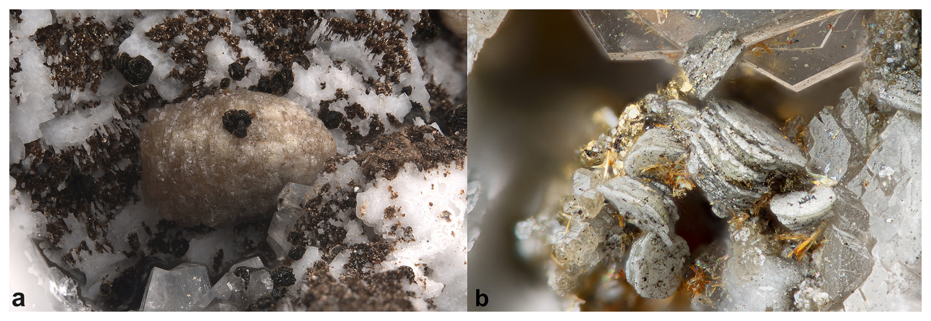
Figure 1Bainbridgeite-(NdCe) from Mont Saint-Hilaire, Quebec, Canada: (a) pale-orange-yellow barrel-shaped prismatic crystal 2.5 mm in size with a bainbridgeite-(YCe) core and a bainbridgeite-(NdCe) rim. Specimen CMNMC 46324 (the holotype of bainbridgeite-(YCe)). (b) Tabular saucer-shaped crystals. FOV 1.1 mm. Specimen CMNMC 52468. Canadian Museum of Nature collection. Photos: François Génier (a) and Michael Bainbridge (b).
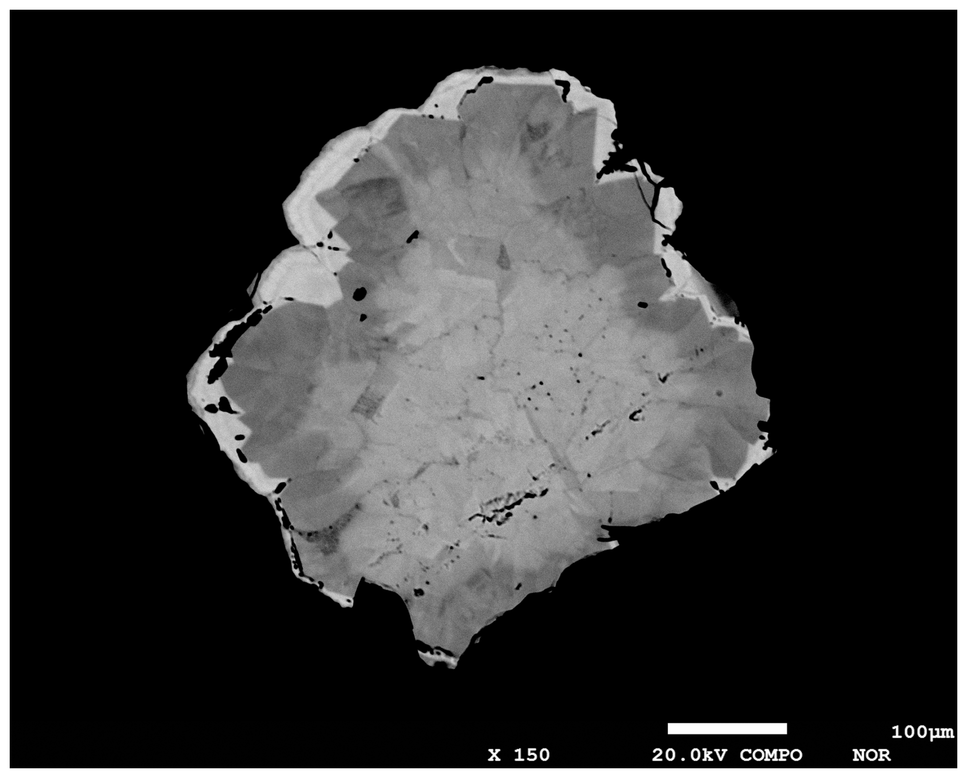
Figure 2Crystal composed of a core of bainbridgeite-(YCe), a darker zone corresponding to an intermediate member of the bainbridgeite-(YCe)–alicewilsonite-(YCe) series, and a bright rim of bainbridgeite-(NdCe). Polished section. SEM (BSE) photo.
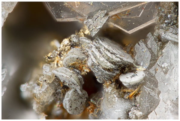
Bainbridgeite-(NdCe) was also found in a hornfels rock together with yellow acicular rutile, colourless hexagonal thin tabular crystals of gmelinite-Na, sphalerite, calcite, analcime, and pyrite. It forms tabular saucer-shaped crystals up to 0.5 mm in size, which are commonly stacked (Fig. 1b). In this case, the entireties of the crystals are bainbridgeite-(NdCe).
Bainbridgeite-(NdCe) is pale yellow, pale orange, white, or pale grey (Fig. 1). The streak is white; the lustre is vitreous. The mineral has no cleavage, and its fracture is uneven. The Mohs hardness of 3 determined on bainbridgeite-(NdCe) from the hornfels rock to be bainbridgeite-(NdCe) zones in the holotype are too thin. The mineral is non-fluorescent under ultraviolet light. The density calculated using the empirical formula of the holotype and unit-cell volume refined from the single-crystal X-ray diffraction data is 3.49 g cm−3.
Bainbridgeite-(NdCe) is optically biaxial (+), α=1.577(3), β=1.592(3), γ=1.657(3), 2V (meas.) = 40(3)° (from a spindle-stage extinction curve), and 2V (calc.) = 52°.
Electron microprobe analyses (EMPAs) of bainbridgeite-(NdCe) were performed using a JEOL 8230 SuperProbe electron microscope equipped with five WDS spectrometers (University of Ottawa – Canadian Museum of Natures MicroAnalysis Laboratory, Canada) using an accelerating voltage of 20 kV, a beam current of 10 nA, and a beam diameter of 10–20 µm depending on the grain size. Bainbridgeite-(NdCe) is unstable under an electron beam; consequently, larger beam diameters were used to minimize element migration. The following reference materials were used: albite or NaInSi2O6 (NaKα), diopside (CaKα), celestine (SrLα), sanbornite (BaLα), YAG (YLα), LaPO4 (LaLα), CePO4 (CeLα), PrPO4 (PrLβ), NdPO4 (NdLα), SmPO4 (SmLα), EuPO4 (EuLα), GdPO4 (GdLα), TbPO4 (TbLα), DyPO4 (DyLβ), HoPO4 (HoLβ), ErPO4 (ErLα), TmPO4 (TmLα), YbPO4 (YbLα), ThO2 (ThMα), and UO2 (UMα). The intensity data were corrected for time-dependent intensity (TDI) loss (or gain) using a self-calibrated correction for NaKα, CaKα, YLα, and LaLα. H2O and CO2 contents were not analysed due to the paucity of the available material. Elemental interferences were corrected using empirical correction factors (Donovan et al., 1993). Raw data were matrix corrected using the Armstrong–Brown–Love–Scott Phi Rho Z correction (Armstrong, 1988).
The Fourier transform infrared (FTIR) spectrum of bainbridgeite-(NdCe) was obtained using a Bruker Hyperion 2000 microscope interfaced to a Tensor 27 spectrometer with a wide-band mercury cadmium telluride (MCT) detector (Canadian Conservation Institute, Canada). A small fragment of bainbridgeite-(NdCe) was mounted on a low-pressure diamond anvil microsample cell and analysed in transmission mode. The spectrum was collected between 4000–400 cm−1 with the co-addition of 150 scans at a 4 cm−1 resolution.
Powder X-ray diffraction (PXRD) data were collected at the Canadian Museum of Nature, Canada, using a Bruker D8 Discover microdiffractometer equipped with a DECTRIS EIGER2 R 500K detector and IµS microfocus X-ray source ( Å), with the Kα2 contribution removed using the “Strip Kα2” tool in Bruker Diffrac.EVA V4.3. The instrument was calibrated using a statistical calibration method (Rowe, 2009). A powder ball ∼100 µm in diameter, mounted on a fibre pin mount, was analysed with continuous Phi rotation and 10° rocking motion along the Psi axis of the centric Eulerian cradle stage. We could not recover enough bainbridgeite-(NdCe) grains for PXRD analysis, and so the obtained pattern represents an average between bainbridgeite-(NdCe) and bainbridgeite-(YCe).
Single-crystal X-ray diffraction (SXRD) studies were carried out at the Natural History Museum, University of Oslo, Norway, using a Rigaku XtaLAB Synergy-S diffractometer equipped with a HyPix 6000HE detector () operating at 50 kV and 1 mA. The data were processed, including an absorption correction, using Rigaku's CrysAlis Pro software. The crystal used to collect electron microprobe data was subsequently sampled for the single-crystals X-ray study. However, because bainbridgeite-(NdCe) occurs as a thin rim with varying thickness on bainbridgeite-(YCe) and because the boundary between the two phases is not visually identifiable, we could not sample a pure bainbridgeite-(NdCe) grain for the SXRD analysis. Therefore, the obtained structural model represents an average between bainbridgeite-(NdCe) and bainbridgeite-(YCe).
Table 1Chemical data for bainbridgeite-(NdCe).
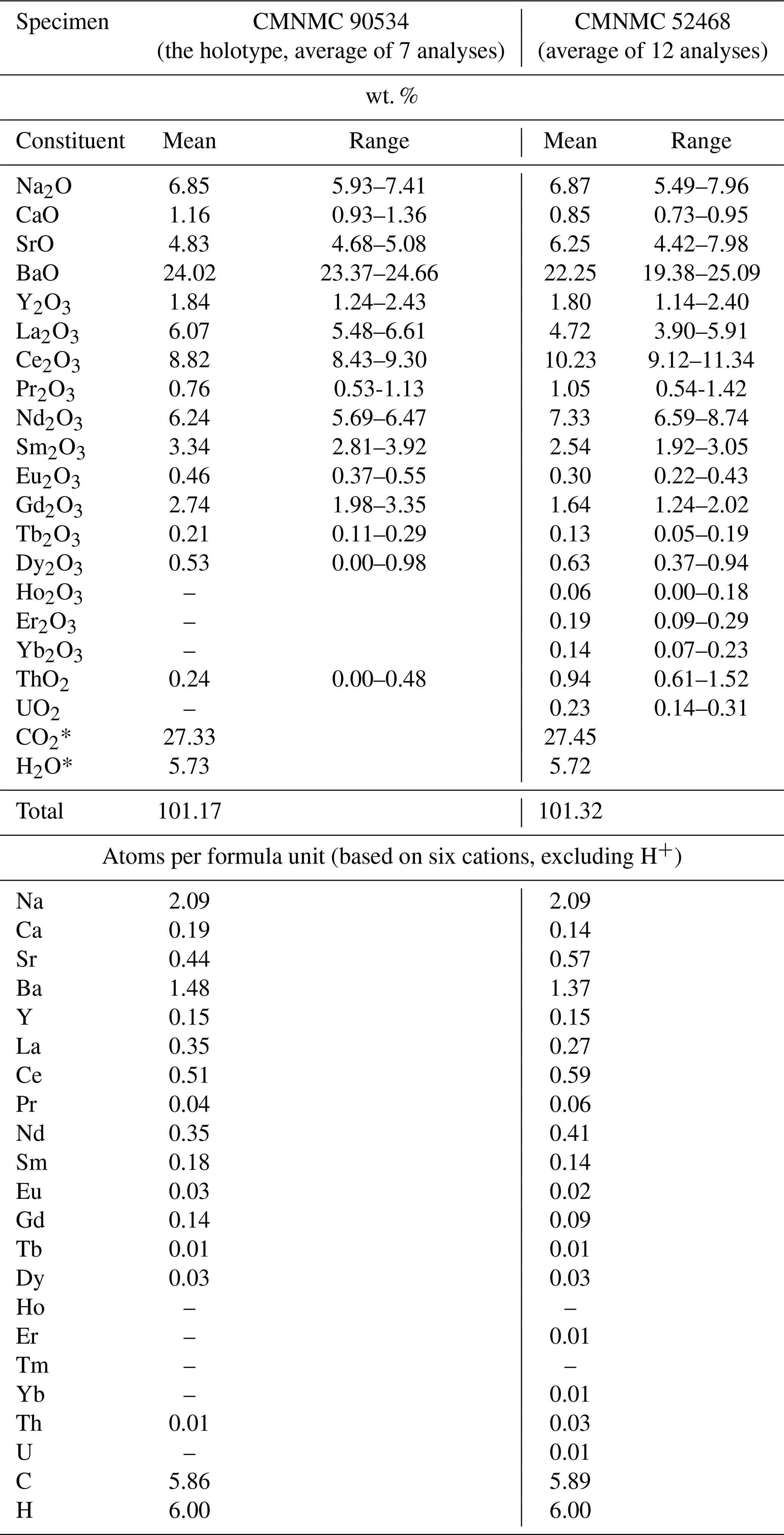
* Calculated from the stoichiometry. Dash means the content of a constituent is below the detection limit.
5.1 Chemical data
Representative EMPAs for bainbridgeite-(NdCe) collected on several specimens from Mont Saint-Hilaire are given in Table 1. The contents of Zr, Tm, Lu, and Hf are below the detection limit.
Atoms per formula unit (apfu) for the holotype, calculated on the basis of six cations, excluding H and C, are as follows: Na2.09Ca0.19Sr0.44Ba1.48Y0.15La0.35Ce0.51Pr0.04Nd0.35Sm0.18
Eu0.03Gd0.14Tb0.01Dy0.03Th0.01C5.86H6O20.4. The ideal end-member formula is Na2Ba2NdCe(CO3)6•3H2O, which requires Na2O 6.08, BaO 30.09, Nd2O3 16.51, Ce2O3 16.11, CO3 25.91, H2O 5.30, total 100 wt. %.
5.2 Infrared spectroscopy
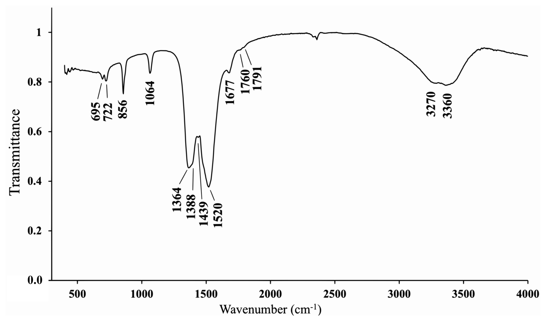
The IR spectrum of bainbridgeite-(NdCe) (Fig. 3) shows IR bands of O–H-stretching (in the range from 3270 to 3360 cm−1) and H–O–H-bending (at 1677 cm−1) vibrations of H2O molecules and C–O-stretching (in the range 1364–1520 cm−1) vibrations of CO-group molecules. The band at 1064 cm−1 can be assigned to the non-degenerate mode of C–O-stretching vibrations, indicating polarization of CO groups, as this mode would have been inactive if symmetric non-polarized carbonate groups (with a three-fold axis) were present in the IR spectrum. The band assignment was made in accordance with Chukanov and Chervonnyi (2016).
Table 2X-ray powder diffraction data (d in Å) for bainbridgeite-(NdCe). The strongest reflections are given in bold.
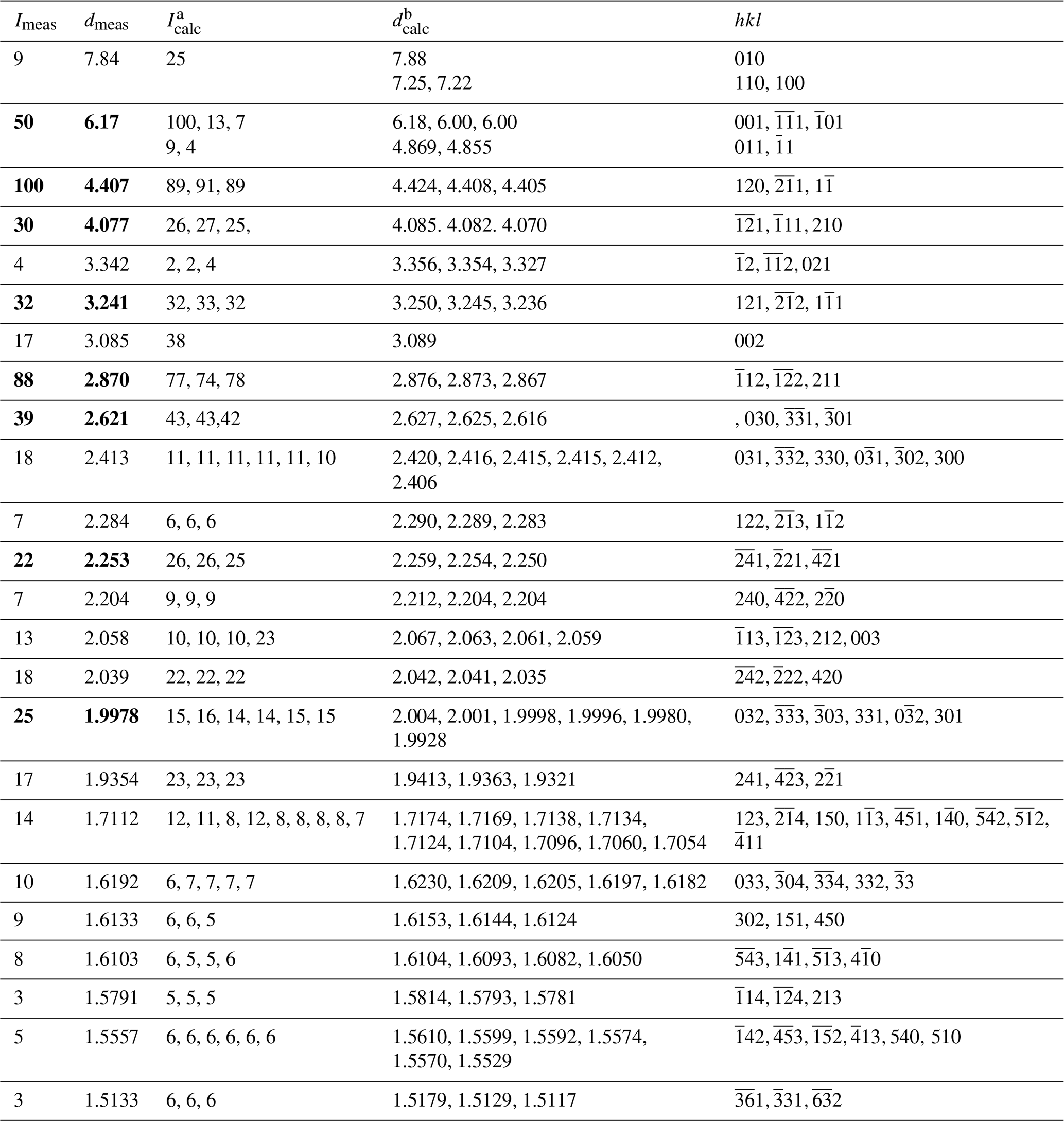
a Calculated from the crystal structure determination; only reflections with intensities >4 are given. b Calculated from PXRD Rietveld unit-cell refinement with a=9.0705(6) Å, b=9.1075(4) Å, c=6.8840(3) Å, α=102.640(8)°, β=116.187(5)°, γ=59.917(7)°, and V=441.57(5) Å3.
5.3 X-ray diffraction data and description of the crystal structure
The indexed PXRD data are given in Table 2. The parameters of the triclinic unit cell refined using Bruker DIFFRAC.TOPAS software are as follows: a=9.0705(6) Å, b=9.1075(4) Å, c=6.8840(3) Å, α=102.640(8)°, β=116.187(5)°, γ=59.917(7)°, and V=441.57(5) Å3.
Table 3Crystal data, data collection information, and structure refinement details for bainbridgeite-(NdCe).
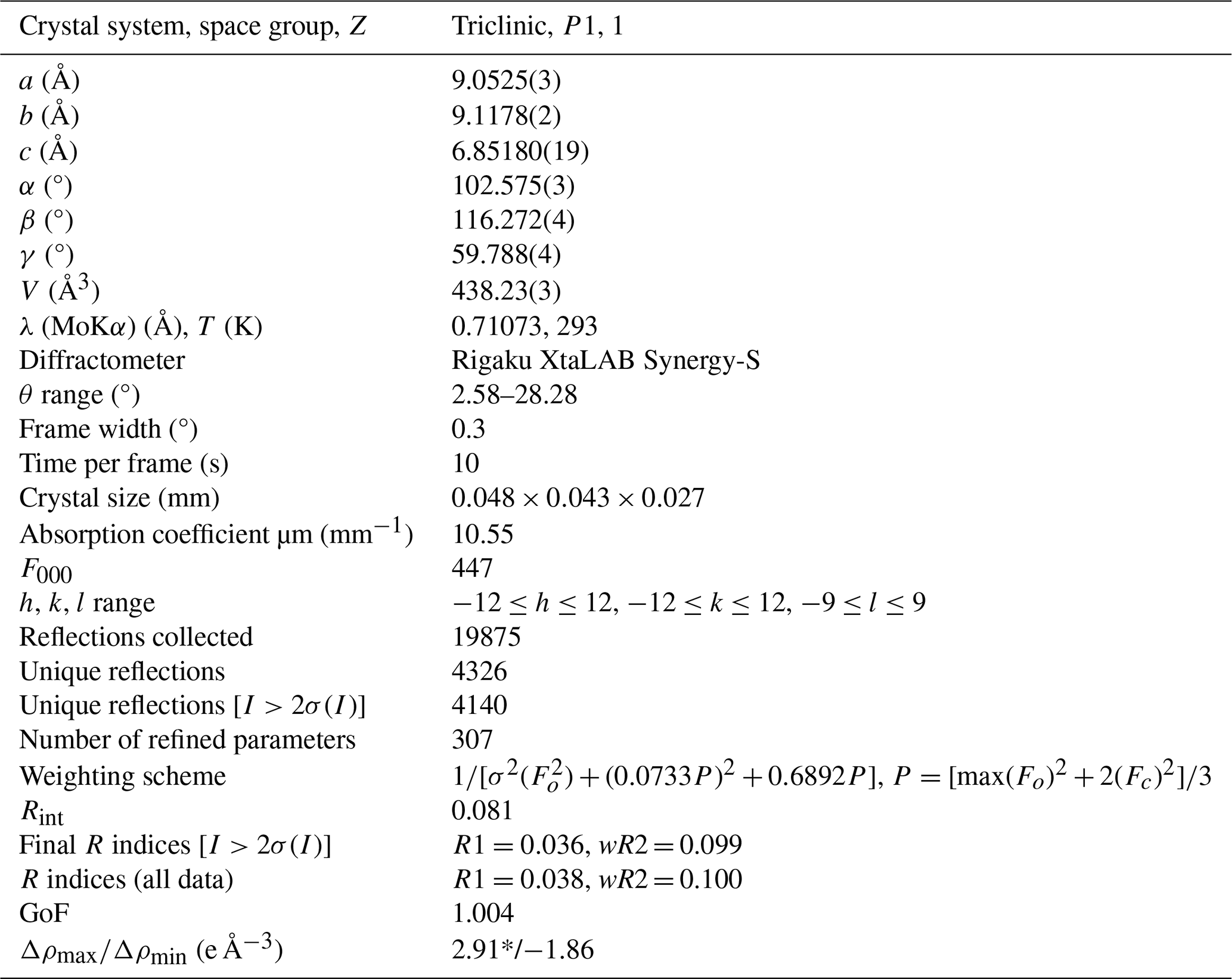
* Located 0.73 Å away from the Ba3 site. There are several peaks up to 2.9 e Å−3 located 0.7–0.8 Å away from large cation sites. A relatively poor overall quality of the SXRD data due to the poor quality of bainbridgeite-(NdCe) crystals resulted in spurious peaks and holes in residual electron density.
Table 4Coordinates and equivalent displacement parameters (Ueq, in Å2) of atoms, site occupancies, and bond valence sums (BVSs) for bainbridgeite-(NdCe).
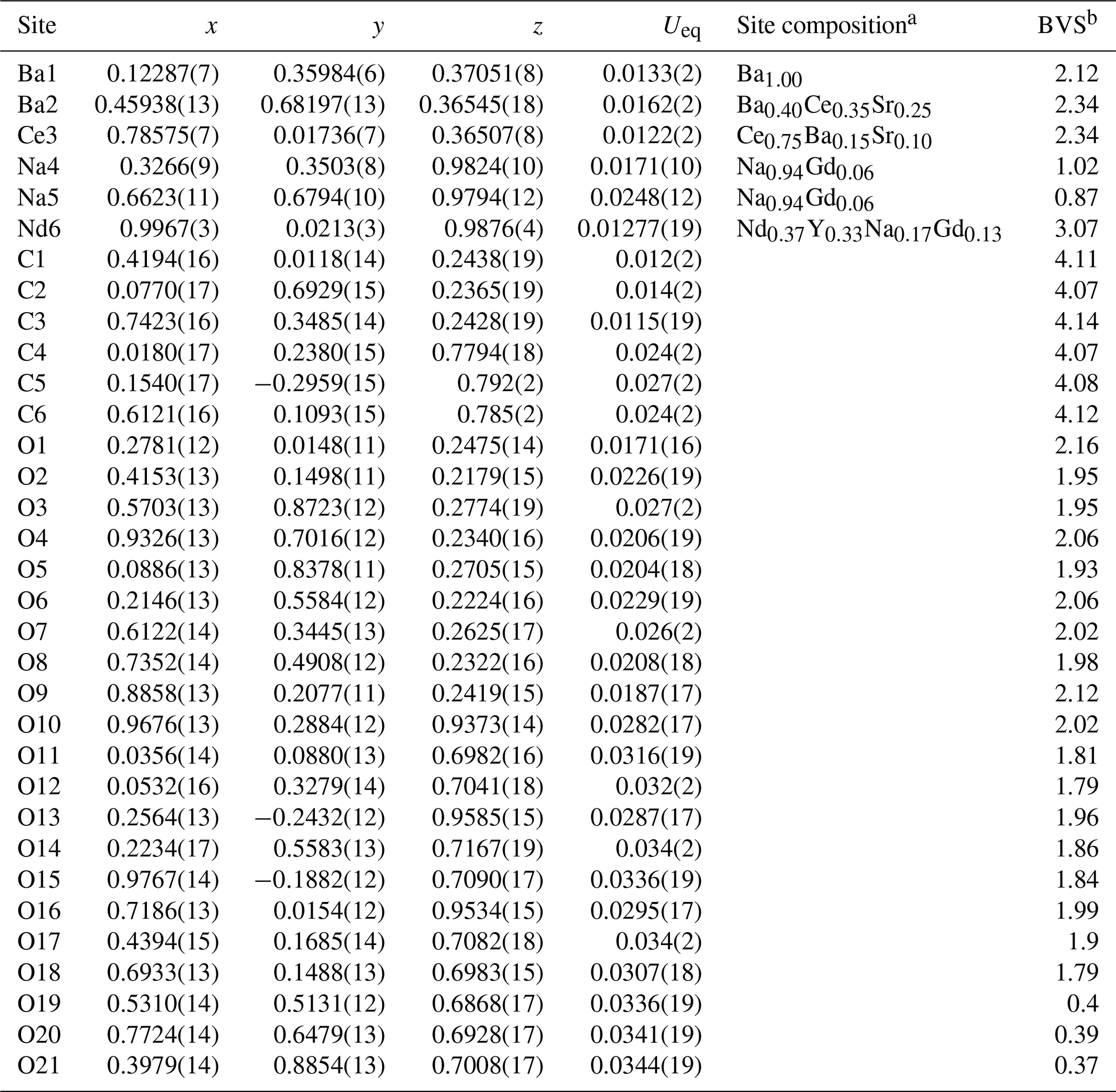
a Based on the eref values, electron microprobe data, interatomic distances, bond valence calculations, and the charge balance. b Bond valence parameters were taken from Gagné and Hawthorne (2015).
Table 5Selected interatomic distances (Å) in the structure of bainbridgeite-(NdCe). The average distances are given in bold.
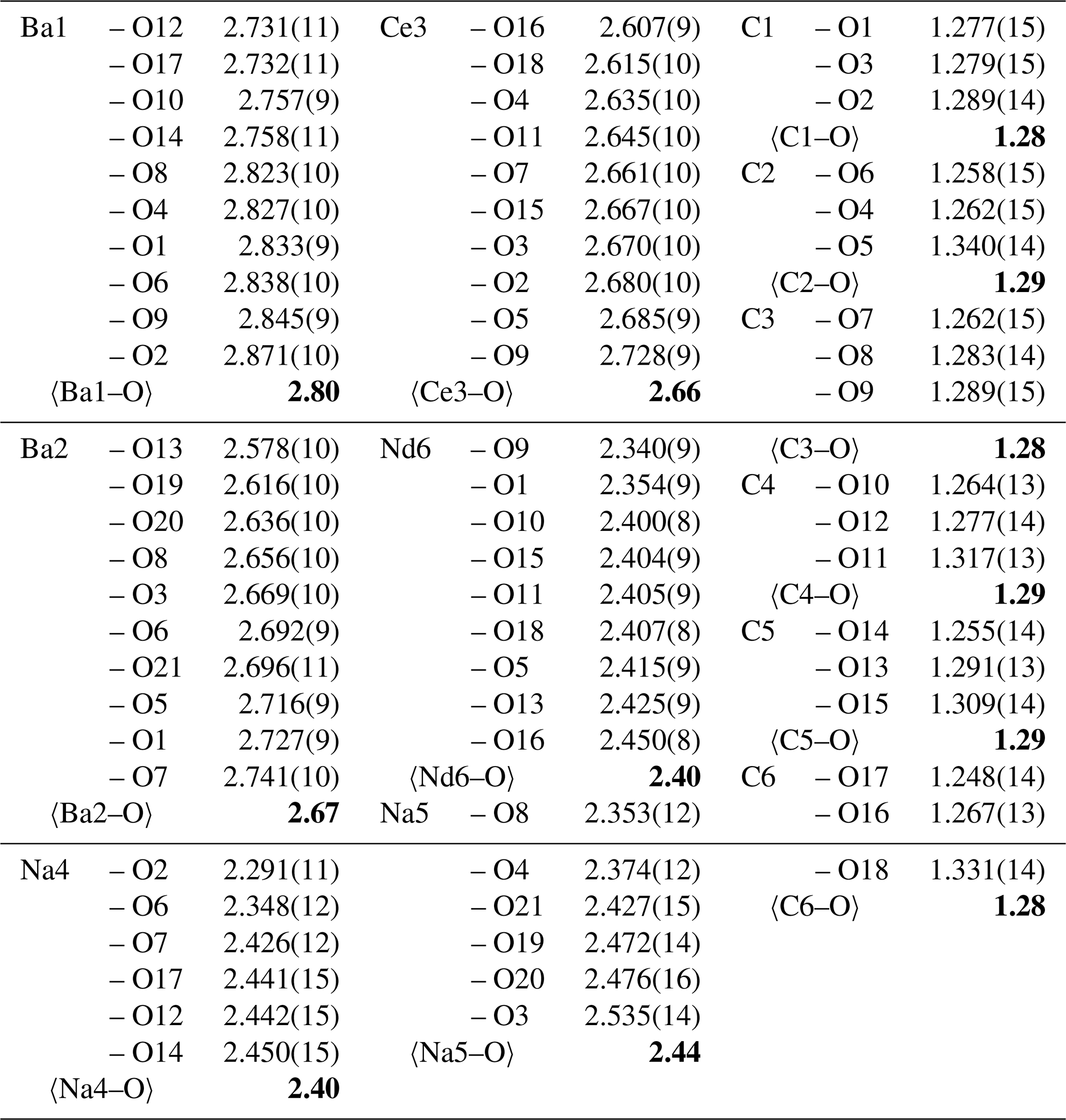
The single-crystal X-ray diffraction data were indexed in the P1 space group with the following unit-cell parameters: a=9.0525(3) Å, b=9.1178(2) Å, c=6.85180(19) Å, α=102.575(3)°, β=116.272(4)°, γ=59.788(4)°, and V=438.23(3) Å3. The structure was solved and refined to R1=0.036 on the basis of 4140 independent reflections with I>2σ(I) using the SHELXL-2018/3 programme package (Sheldrick, 2015). Crystal data, data collection information, and structure refinement details are given in Table 3; atom coordinates, equivalent displacement parameters, site composition, and bond valence sums (BVSs) are given in Table 4; and selected interatomic distances are given in Table 5. The crystallographic information file (CIF) for bainbridgeite-(NdCe) is available as a Supplementary file. It was also deposited in the Inorganic Crystal Structure Database (ICSD; #CSD 2555428).
SXRD studies of the mckelveyite group minerals are challenging because of the poor quality of their crystals, resulting in multiple split reflections and streaks. Although the quality of the bainbridgeite-(NdCe) crystal was good for the minerals in the group, it was still relatively poor overall.
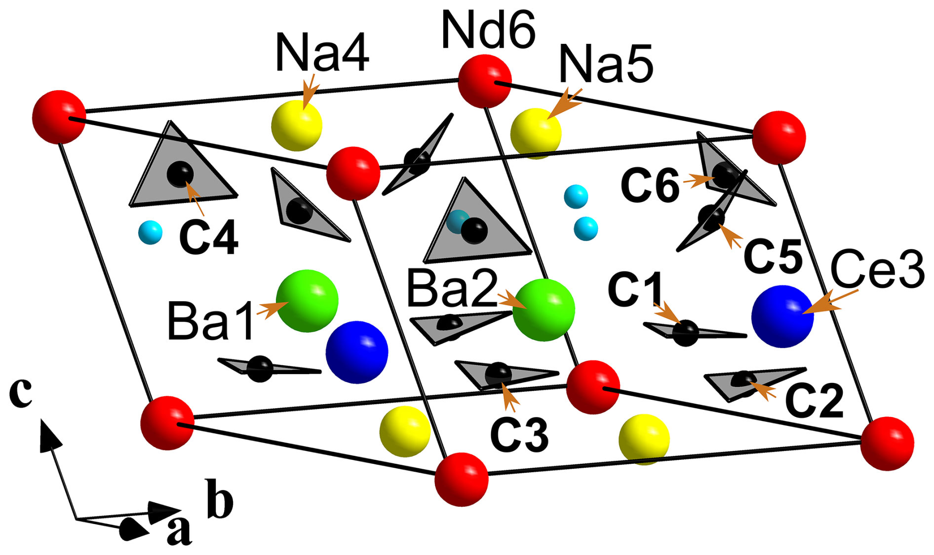
Figure 4General view of the crystal structure of bainbridgeite-(NdCe). Sky-blue spheres are H2O molecules. The unit cell is outlined.
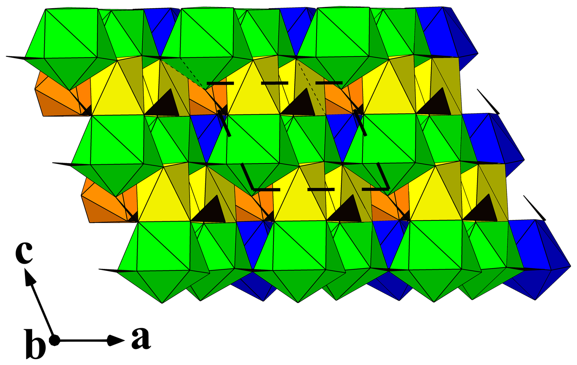
Figure 5A view along [010] of the crystal structure of bainbridgeite-(NdCe). Ba-, Ce-, Nd-, and Na-centred polyhedra are shown in light green, blue, orange, and yellow, respectively. Carbonate groups are black triangles. The unit cell is outlined.
Bainbridgeite-(NdCe) Na2Ba2NdCe(CO3)6•3H2O is strongly pseudotrigonal. There are six independent large cation sites in the structure (Fig. 4) forming two alternating layers parallel to the ab plane (Fig. 5). The Na, Ba, Sr, Ce, Nd, and Gd atoms were distributed among these sites based on the microprobe data, refined site-scattering factors (eref, in electrons per site), and charge balance, taking into account bond valence sums (BVS) and interatomic distances (Tables 4–5). The Ca atoms were not included in the refinement; lanthanoids (Ln, La–Lu) lighter than Nd were formally refined as Ce atoms, and Ln heavier than Nd were formally refined as Gd atoms. One of the layers is formed by the Ba1, Ba2, and Ce3 sites that have 10-fold coordination. The large Ba1-centred polyhedron with the 〈Ba1–O〉 distance of 2.80 Å is occupied by Ba atoms. The refinement showed that the remaining Ba, Ce, and Sr atoms are distributed between Ba2 and Ce3 sites. Their occupancies were refined as Ba0.701(8)Sr0.299(8) (eref=50.6) for the Ba2 site and Ce0.897(9Sr0.103(9) (eref=56.0) for the Ce3 site. Sr atoms prefer the Ba2-centred polyhedron; thus, the Ce3-centred polyhedron with the 〈Ce3–O〉 distance of 2.68 Å should be occupied predominantly by Ce atoms as it is too small for Ba atoms. The preference of Ce atoms for the Ce3 site was also observed in alicewilsonite-(YCe) (Lykova et al., 2023b). The assigned occupancies are Ba0.40Ce0.35Sr0.25 and Ce0.75Ba0.15Sr0.10 for the Ba2 and Ce3 sites, respectively. Na4 and Na5 sites have octahedral coordination, and their occupancies were fixed as Na0.94Gd0.06. The occupancy for the Nd6 site was refined as Nd0.466(8)Y0.365(8)Na0.17 (eref=44.4) and fixed as Nd0.37Y0.33Na0.17Gd0.13.
There are six carbonate groups. The three CO groups centred by carbon atoms C1, C2, and C3 are almost coplanar with {001}, while the other three centred by C4, C5, and C6 atoms are not coplanar (Fig. 4).
The BVSs at the O19, O20, and O21 sites (0.40, 0.39, and 0.37 valence units, respectively) indicate the presence of H2O molecules, also confirmed by the presence of the bands of O–H-stretching and H–O–H bending vibrations in the IR spectrum of bainbridgeite-(NdCe) (Fig. 3). The three H2O molecules are bonded to Ba2- and Na5-centred polyhedra that share a face.
The structural formula derived from the refinement is
(Na1.88Gd0.12)∑2.00(Ba1.40Ce0.35Sr0.25)∑2.00(Nd0.37Y0.33
Na0.17Dy0.13)∑1.00(Ce0.75Ba0.15Sr0.10)∑1.00(CO3)6(H2O)3, which leads to the ideal formula Na2Ba2NdCe(CO3)6•3H2O.
Table 6Refined site-scattering factors and assigned occupancies for Ba2, Ca3, and Nd/Y6 sites in the structures of bainbridgeite-(NdCe) (upper lines) and bainbridgeite-(YCe) (lower lines, after Lykova et al., 2024).
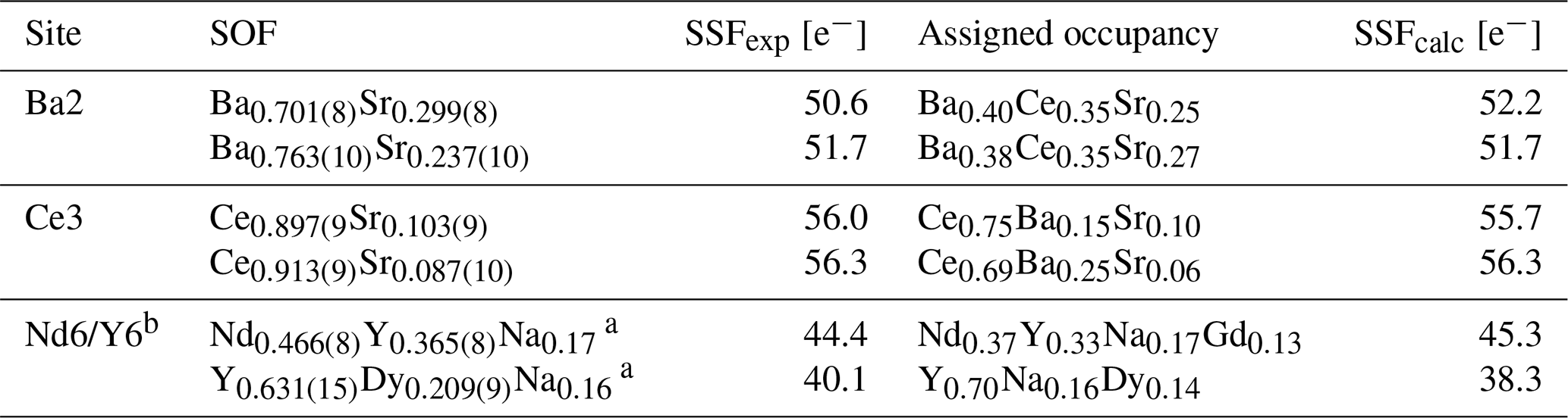
SOF: refined site occupation factor; SSFexp and SSFcalc: experimental and calculated site-scattering factors (numbers of electrons). a The occupancies of Na were fixed. b In the structure of bainbridgeite-(NdCe) and bainbridgeite-(YCe), respectively.
Bainbridgeite-(NdCe) Na2Ba2NdCe(CO3)6•3H2O is a member of the mckelveyite group (Lykova et al., 2023c). It is the Nd analogue of bainbridgeite-(YCe) Na2Ba2YCe(CO3)6•3H2O (Lykova et al., 2024) and the BaNd analogue of alicewilsonite-(YCe) Na2Sr2YCe(CO3)6•3H2O (Lykova et al., 2023b). It is closely related to donnayite-(Y) NaCaSr3Y(CO3)6•3H2O (Chao et al., 1978) and mckelveyite-(Y) NaCaBa3Y(CO3)6•3H2O (Milton et al., 1965, Chao et al., 1978).
The separation and concentration of Ce and Nd atoms at two different sites of the structure are unique and highly unusual features of the described phase and thus require rigorous examination to prove that the proposed distribution is conclusive.
Neodymium content in the holotype of bainbridgeite-(NdCe) (0.35 apfu) is
-
significantly higher than the content of any other individual, heavier Ln (e.g. 0.18 apfu Sm),
-
compatible with the total of all the other heavier Ln (Sm–Dy, 0.39 apfu),
-
significantly higher than the content of Y (0.15 apfu),
-
significantly higher than the content of Nd in the holotype of bainbridgeite-(YCe) (0.11 apfu, Lykova et al., 2024).
Bainbridgeite-(NdCe) from the hornfels rock (specimen CMNMC 52468; Table 1) shows a similar pattern with an even higher content of Nd (0.41 apfu), a comparable content of Y (0.15 apfu), and a lower total of the heavier Ln (Sm-Yb, 0.31 apfu).
In both finds the content of lighter Ln (La–Pr; 0.90 and 0.92 apfu, respectively) is close to the expected value for bainbridgeite – 1 apfu.
Therefore, even when anticipating the distribution of some Nd atoms among other cations sites in bainbridgeite-(NdCe), most atoms must be concentrated at the Nd, which makes it the dominant – and, thus, the species-defining – cation for the species based on the chemical data only.
Structural evidence also strongly points to the preferential entry of Nd atoms at the Nd6 site. Different parts of crystals (rims and cores) from the same find were used to solve the structure of both bainbridgeite-(NdCe) and bainbridgeite-(YCe).
The distribution of cations between the sites in the two minerals is very similar (Table 6) except for the Nd6/Y6 site. The latter is occupied predominantly by Y atoms (70 %) in bainbridgeite-(YCe) and by heavier Ln atoms in bainbridgeite-(NdCe), indicating that the entry of heavier Ln atoms follows the substitution scheme Y Ln3+ at the Nd6/Y6 site. This can be corroborated with the significant negative correlation (R2=0.82) between Y and heavier Ln contents we described previously in bainbridgeite-(YCe) (Lykova et al., 2024).
Note that the Y content in bainbridgeite-(NdCe) based on the structural analysis (0.33 atoms per formula unit [apfu]) is significantly higher than the value obtained from electron microprobe data (0.15 apfu) due to the presence of both bainbridgeite-(NdCe) and bainbridgeite-(YCe) in the grain used for the SXRD study, as discussed in Sect. 4.
The empirical formula of the holotype of bainbridgeite-(NdCe), written taking into account the structural data, is
(Na1.92HREE0.08)∑2.00(Ba1.34Sr0.33Ca0.18LREE0.15)∑2.00
(Nd0.35HREE0.31Na0.17Y0.15Th0.01Ca0.01)∑1.00(LREE0.75
Ba0.14Sr0.11)∑1.00(CO3)5.86(H2O)3.00, where LREE = (Ce0.51La0.35Pr0.04)∑0.90, and HREE = (Sm0.18Gd0.14Eu0.03Dy0.03Tb0.01)∑0.39.
For comparison, the empirical formula of the holotype of bainbridgeite-(YCe) is
(Na1.95HREE0.05)∑2.00(Ba1.33LREE0.30Sr0.27Ca0.10)∑2.00
(Y0.70Na0.16HREE0.14)∑1.00(LREE0.63Ba0.27Sr0.10)∑1.00
(CO3)5.86(H2O)3.00, where LREE = (Ce0.49La0.29Nd0.11Pr0.03Sm0.01)∑0.93, and HREE = (Dy0.07Er0.05Gd0.03Ho0.02Yb0.02)∑0.19 (Lykova et al., 2024).
The ability of mckelveyite-group minerals to selectively concentrate Nd but not lighter rare-earth elements (REEs) at the Nd6/Y6 site was also observed in mckelveyite-(Nd) (Lykova et al., 2023a).
All the above observations point to the same preferential concentration of Nd atoms as the Nd6/Y6 site.
Generally, neodymium disperses in both Ce- and Y-dominant minerals and rarely forms its own phases. We showed, using the example of piilonenite-(Nd), Nd(CO3)2•3H2O (Lykova et al., 2026), another Nd-dominant carbonate from Mont Saint-Hilaire, that neodymium can separate and concentrate in late-stage agpaitic environments due to its crystal chemically driven affinity for yttrium. With the depletion of Y from a late-stage solution, Nd can become the prevalent rare-earth element (REE) in a phase. We propose that the formation of bainbridgeite-(NdCe) followed the same mechanism.
Under this model we should expect samarium and gadolinium cations, which are smaller than neodymium but larger than heavier REEs that usually concentrate in Y-dominant minerals Y (Dy, Yb), to show an even greater affinity for yttrium and thus also concentrate at Y-favouring sites in mineral structures in late-stage processes. The chemical data show that contents of Sm and Gd atoms are, respectively,
-
0.01 and 0.03 apfu in the holotype of bainbridgeite-(YCe) from the “carbonate pegmatite” (cores of the crystals; Lykova et al., 2024),
-
0.18 and 0.14 apfu in the holotype of bainbridgeite-(NdCe) from the same pegmatite (rims of the crystals),
-
0.14 and 0.09 apfu in bainbridgeite-(NdCe) from the hornfels rock.
As predicted, bainbridgeite-(NdCe) shows a significant enrichment in both Sm and Gd. Piilonenite-(Nd) is also characterized by significant contents of Sm and Gd atoms but not heavier REEs (Lykova et al., 2026).
Crystallographic data for bainbridgeite-(NdCe) are available in the Supplement.
The supplement related to this article is available online at https://doi.org/10.5194/ejm-38-419-2026-supplement.
IL conceptualized the project. RR collected the powder X-ray diffraction data and sub-sampled the specimens. EMPAs were obtained by GP. HF collected the single-crystal X-ray diffraction data. KO measured the optical properties. SB obtained the IR spectrum. IL processed the data and interpreted the results. The paper was written by IL with contributions from all of the co-authors.
The contact author has declared that none of the authors has any competing interests.
Publisher's note: Copernicus Publications remains neutral with regard to jurisdictional claims made in the text, published maps, institutional affiliations, or any other geographical representation in this paper. The authors bear the ultimate responsibility for providing appropriate place names. Views expressed in the text are those of the authors and do not necessarily reflect the views of the publisher.
We would like to thank two anonymous referees for the critical reading, Elsa Pfenninger-Horváth and László Horváth for providing us with the specimens for the study, and François Génier and Michael Bainbridge for taking the colour photos.
This research was financially supported by the Canadian Museum of Nature.
This paper was edited by Sergey Krivovichev and reviewed by two anonymous referees.
Armstrong, J. T.: Quantitative analysis of silicate and oxide minerals: comparison of Monte Carlo, ZAF and phi-rho-z procedures, Analysis Microbeam, 239–246, 1988.
Bayliss, P. and Levinson, A. A.: A system of nomenclature for rare-earth mineral species; revision and extension, Am. Mineral., 73, 422–423, 1988.
Chao, G. Y., Harris, D. C., Hounslow, A. W., Mandarino, J. A., and Perrault, G.: Minerals from the nepheline syenite, Mont Saint Hilaire, Quebec, Can. Mineral., 9, 109–123, 1967.
Chao, G. Y., Mainwaring, P. R., and Baker, J.: Donnayite, NaCaSr3Y(CO3)6•3H2O, a new mineral from Mont St-Hilaire, Quebec, Can. Mineral., 16, 335–340, 1978.
Chukanov, N. V. and Chervonnyi, A. D.: Infrared Spectroscopy of Minerals and Related Compounds, Springer Cham, Switzerland, 1109 pp., https://doi.org/10.1007/978-3-319-25349-7, 2016.
Donovan, J. J., Snyder, D. A., and Rivers, M. L.: An improved interference correction for trace element analysis, Microbeam Analysis, 2, 23–28, 1993.
Gagné, O. C. and Hawthorne, F. C.: Comprehensive derivation of bond-valence parameters for ion pairs involving oxygen, Acta Crystallogr. B: Struct. Sci. Cryst. Eng. Mater., 71, 562–578, 10.1107/s2052520615016297, 2015.
Horváth, L., Gault, R. A., Pfenninger-Horvath, E., and Poirier, G.: Mont Saint-Hilaire: History, Geology, Mineralogy, The Canadian Mineralogist Special Publication 14, Mineralogical Association of Canada, Canada, 634 pp. 2019.
Levinson, A. A.: A system of nomenclature for rare-earth minerals, Am. Mineral., 51, 152–158, 1966.
Lykova, I., Rowe, R., Poirier, G., and Barnes, S.: Mckelveyite-(Nd), IMA 2023-017, in: CNMNC Newsletter 74, Eur. J. Mineral., 35, https://doi.org/10.5194/ejm-35-659-2023, 2023a.
Lykova, I., Rowe, R., Poirier, G., Friis, H., and Helwig, K.: Mckelveyite group minerals – Part 2: Alicewilsonite-(YCe), Na2Sr2YCe(CO3)6⋅3H2O, a new species, Eur. J. Mineral., 35, 143–155, https://doi.org/10.5194/ejm-35-143-2023, 2023b.
Lykova, I., Rowe, R., Poirier, G., Giester, G., Ojaste, K., and Friis, H.: Mckelveyite group minerals – Part 1: Nomenclature and new data on donnayite-(Y), Eur. J. Mineral., 35, 133–142, https://doi.org/10.5194/ejm-35-133-2023, 2023c.
Lykova, I., Rowe, R., Poirier, G., Friis, H., and Helwig, K.: Mckelveyite group minerals – Part 3: Bainbridgeite-(YCe), Na2Ba2YCe(CO3)6⋅3H2O, a new species from Mont Saint-Hilaire, Canada, Eur. J. Mineral., 36, 183–194, https://doi.org/10.5194/ejm-36-183-2024, 2024.
Lykova, I., Rowe, R., Teat, S. J., Poirier, G., and Barnes, S.: Piilonenite-(Nd), NaNd(CO3)2⋅3H2O, a new neodymium-dominant carbonate mineral from Mont Saint-Hilaire, Quebec, Canada, Eur. J. Mineral., 38, 337–345, https://doi.org/10.5194/ejm-38-337-2026, 2026.
Milton, C., Ingram, B., Clark, J. R., and Dwornik, E. J.: Mckelveyite, a new hydrous sodium barium rare-earth uranium carbonate mineral from The Green River Formation, Wyoming, Am. Mineral., 50, 593–612, 1965.
Normand, C. and Tarassoff, P.: Mineralogy and geology of the Poudrette quarry, Mont Saint-Hilaire, Geological Association of Canada – Mineralogical Association of Canada, Guidebook, Field trip A4, 23 pp. 2006.
Rowe, R.: New statistical calibration approach for Bruker AXS D8 Discover microdiffractometer with Hi-Star detector using GADDS software, Powder Diffraction, 24, 263–271, https://doi.org/10.1154/1.3193683, 2009.
Sheldrick, G. M.: Crystal structure refinement with SHELXL, Acta Crystallogr. C Struct. Chem., 71, 3–8, 10.1107/s2053229614024218, 2015.