the Creative Commons Attribution 4.0 License.
the Creative Commons Attribution 4.0 License.
 | 12 Aug 2020
Luxembourgite, AgCuPbBi4Se8, a new mineral species from Bivels, Grand Duchy of Luxembourg
Simon Philippo
Yannick Bruni
Pietro Vignola
Jiří Sejkora
Luxembourgite, ideally AgCuPbBi4Se8, is a new selenide discovered at Bivels, Grand Duchy of Luxembourg. The mineral forms tiny fibres reaching 200 µm in length and 5 µm in diameter, which are deposited on dolomite crystals. Luxembourgite is grey, with a metallic lustre and without cleavage planes; its Mohs hardness is 3 and its calculated density is 8.00 g cm−3. Electron-microprobe analyses indicate an empirical formula Ag1.00(Cu0.82Ag0.20Fe0.01)Σ1.03Pb1.13Bi4.11(Se7.72S0.01)Σ7.73, calculated on the basis of 15 atoms per formula unit. A single-crystal structure refinement was performed to R1=0.0476, in the P21∕m space group, with a=13.002(1), b=4.1543(3), c=15.312(2) Å, , V=782.4(2) Å3, Z=2. The crystal structure is similar to that of litochlebite and watkinsonite and can be described as an alternation of two types of anionic layers: a pseudotetragonal layer four atoms thick and a pseudohexagonal layer that is one atom thick. In the pseudotetragonal layers the Bi1, Bi2 ,Bi3, Pb, and Ag1 atoms are localised, while the Cu2 and Bi4 atoms occur between the pseudotetragonal and the pseudohexagonal layers. Bi1, Bi2, and Bi3 atoms occur in weakly distorted octahedral sites, whereas Bi4 occurs in a distorted 7-coordinated site. Ag1 occupies a fairly regular octahedral site, Cu2 a tetrahedral position, and Pb occurs on a very distorted 8-coordinated site.
- Article
(4093 KB) - Full-text XML
-
Supplement
(67 KB) - BibTeX
- EndNote


The term “Luxembourg” designates a natural region composed of the Grand Duchy of Luxembourg and by the Luxembourg Province in the South of Belgium. The Ardennes mountains are in the north of this region and are named “Oesling” in the Grand Duchy. This massif is constituted of Lower Devonian rocks, mainly schists and quartzites, which were affected by the Variscan metamorphism. Pressures and temperatures were estimated to be 500 ∘C and 3–4 kbar in the region of Libramont, but the metamorphic conditions progressively decrease to the east, reaching 400 ∘C and 2 kbar at Bastogne (Beugnies, 1986; Theye and Fransolet, 1993; Hatert, 2005).
During winter 2012, excavation works were realised by the “Société électrique de l'Our” at Bivels, in the north of the Grand Duchy, exposing some fragments of reddish schists containing mineralised veins. The veins were constituted by translucent dolomite crystals reaching 2 mm in diameter, as well as by siderite. A careful examination under the binocular microscope also showed needles of a grey metallic mineral, with a habit similar to that of millerite. Preliminary Energy-Dispersive X-ray Spectrometry (EDS) measurements indicated that these fibres contained Cu, Bi, Ag, Pb, and Se as essential constituents, thus corresponding to an intermediate between litochlebite, Ag2PbBi4Se8 (Sejkora et al., 2011) and watkinsonite, Cu2PbBi4Se8 (Topa et al., 2010).
Electron-microprobe analyses indicated a Cu ∕ Ag ratio very close to 1.0 in the mineral, in agreement with the formula AgCuPbBi4Se8. A single-crystal X-ray diffraction study showed a crystal structure similar to that of watkinsonite but with significant differences compared to litochlebite. Moreover, the ordering of Cu and Ag at two different crystallographic sites indicated that the mineral was not only an intermediate member between litochlebite and watkinsonite but a separate mineral species. This assumption was confirmed by the very limited solid solution between the new species and litochlebite.
The mineral was named luxembourgite for the city of Luxembourg, close to the locality where it was discovered; it is the first new species found in the Grand Duchy of Luxembourg. The new mineral and its name were approved by the IMA-CNMNC under the number IMA 2018-154. A part of the type sample used for electron-microprobe analyses is stored in the collection of the Natural History Museum of Luxembourg (catalogue number FD040). Another part of the type used for the single-crystal structure determination is stored in the collection of the Laboratory of Mineralogy, University of Liège, catalogue number 21302. A detailed mineralogical characterisation of this new species is given in the present paper.
Luxembourgite was found on dumps produced by the construction of a tunnel by the “Société Electrique de l'Our”, at Bivels, north of the Grand Duchy of Luxembourg (coordinates: 49∘57′8′′ N, 6∘10′41′′ E). Associated minerals are dolomite and siderite. The mineral occurs in red schists containing veins of finely crystallised dolomite and siderite. The dolomite crystals reach 2 mm in diameter and sometimes show tiny fibres of luxembourgite. The veins were formed by a hydrothermal process, which is also responsible for other famous sulfide mineralisations in the area, such as the copper mine of Stolzembourg (Philippo et al., 2007) and the antimony mine of Goesdorf (Philippo and Hanson, 2007; Philippo and Hatert, 2018).
Luxembourgite forms tiny fibres deposited on dolomite crystals; the fibres reach 200 µm in length but only 5 µm in diameter (Fig. 1). The colour is grey, the lustre is metallic, and the streak is black. No cleavage has been observed, and the tenacity is brittle. The Mohs hardness, estimated by comparison with litochlebite (Sejkora et al., 2011), is 3. The density, calculated from the chemical analysis and the single-crystal unit cell parameters, is 8.00 g cm−3. Optical properties were not determined due to small grain size.
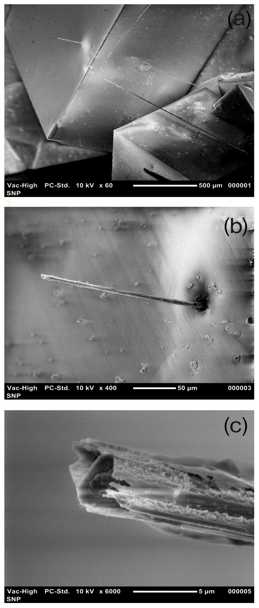
Figure 1Scanning Electron Microscope (SEM) images of luxembourgite fibres occurring on rhombohedral dolomite crystals.
Table 1Electron-microprobe analysis of luxembourgite.
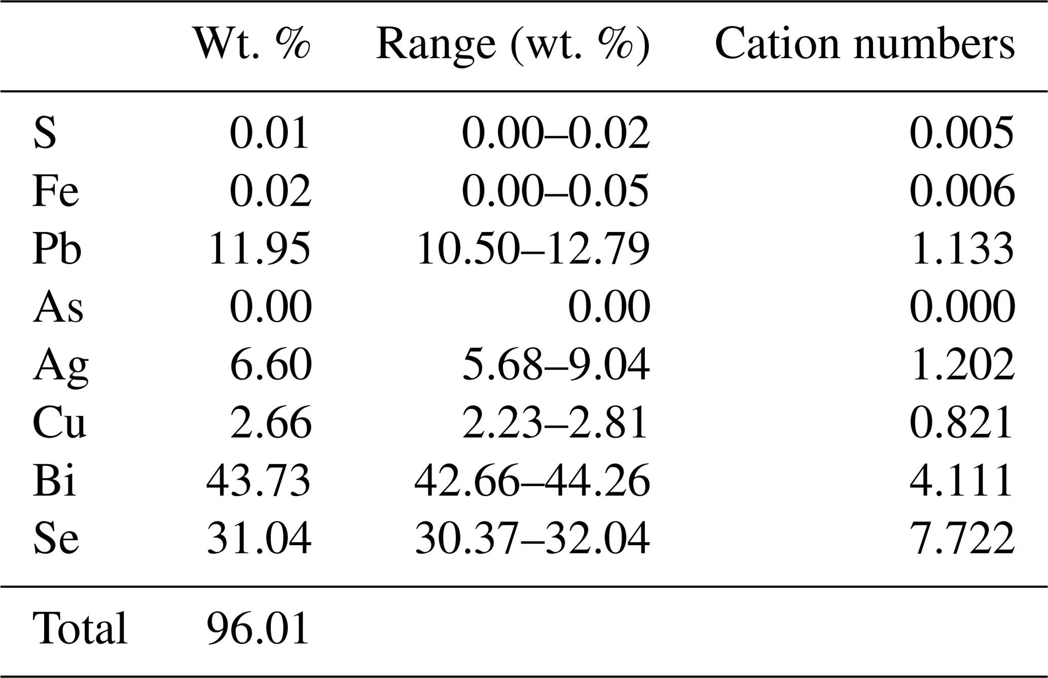
Average of five-point analyses. Cation numbers were calculated on the basis of 15 atoms per formula unit.
Electron-microprobe analyses of luxembourgite (Table 1) were performed with a Jeol JXA-8200 WDS instrument located in Milan, Italy. Acceleration voltage was 15 kV, with a probe current of 5 nA and a beam diameter of 1 µm. Standards used were galena (Pb, S), nickeline (As), and pure metals (Fe, Ag, Cu, Bi, Se). The empirical formula, calculated on the basis of 15 atoms per formula unit, is Ag1.00(Cu0.82Ag0.20Fe0.01)Σ1.03Pb1.13Bi4.11(Se7.72S0.01)Σ7.73. The ideal formula is AgCuPbBi4Se8, which requires Ag 5.58, Cu 3.44, Pb 11.22, Bi 45.28, and Se 34.22 for a total of 100.00 wt %.
Table 2X-ray powder diffraction pattern of luxembourgite (d in Å).
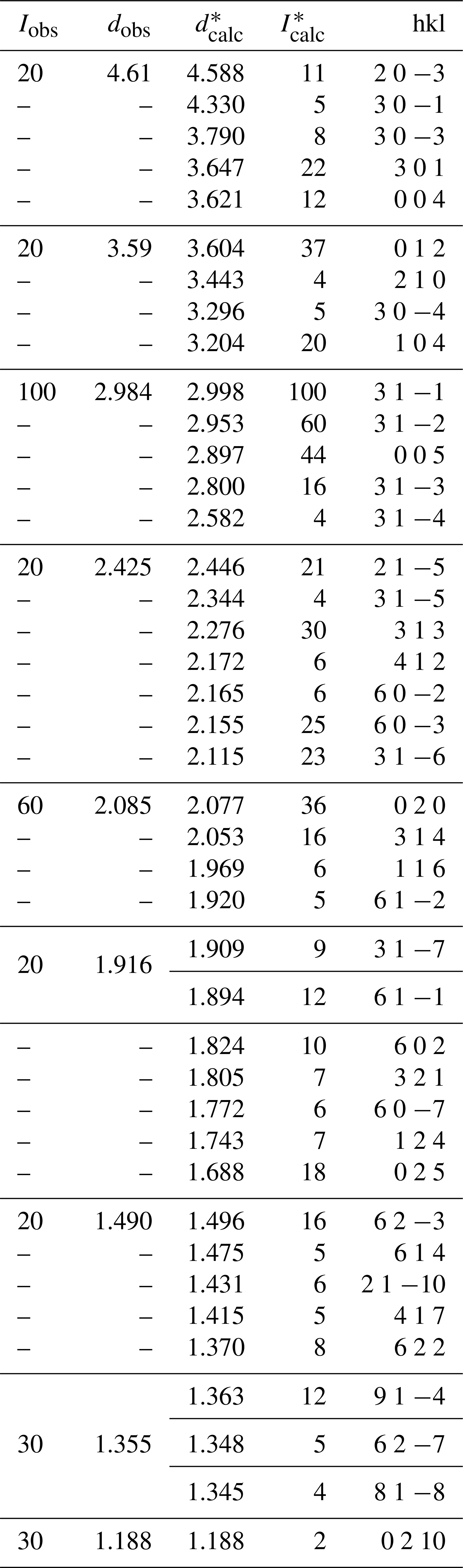
Data collected with a Rigaku Xcalibur four-circle diffractometer, MoKα radiation (λ=0.71073 Å), EOS Rigaku detector. Intensities were estimated visually. Calculated intensities were obtained from the structural data with LAZY PULVERIX (Yvon et al., 1977). Calculated d values were refined with LCLSQ (Burnham, 1962); the refined unit cell parameters are a=13.01(13), b=4.15(2), c=15.36(14) Å, (space group P21∕m). * Only lines with a calculated intensity higher than 4 rel. % are shown.
X-ray powder diffraction data of luxembourgite (Table 2), as well as X-ray single-crystal data, were collected on a Rigaku Xcalibur four-circle diffractometer using the MoKα radiation (λ=0.71073 Å) and equipped with an EOS Charge-Coupled Device (CCD) detector. Unit cell parameters were refined from the powder diffraction data, using the LCLSQ software (Burnham, 1962): a=13.01(13), b=4.15(2), c=15.36(14) Å, , V=783(16) Å3, Z=2, space group P21∕m.
The X-ray structural study was carried out on a fibre of luxembourgite measuring mm. A total of 361 frames with a spatial resolution of 1∘ were collected by the φ∕ω scan technique, with a counting time of 20 s per frame, in the range . A total of 5904 reflections were extracted from these frames, corresponding to 2117 unique reflections. The unit cell parameters refined from these reflections are in very good agreement with those refined from the X-ray powder data (see above). Data were corrected for Lorentz, polarisation and absorption effects, the latter with an empirical method using the SCALE3 ABSPACK scaling algorithm included in the CrysAlisRED package (Oxford Diffraction, 2007). More details on the single-crystal structure refinement are given in Table 3.
Table 3Experimental details for the single-crystal X-ray diffraction study of luxembourgite from Bivels, Luxembourg.
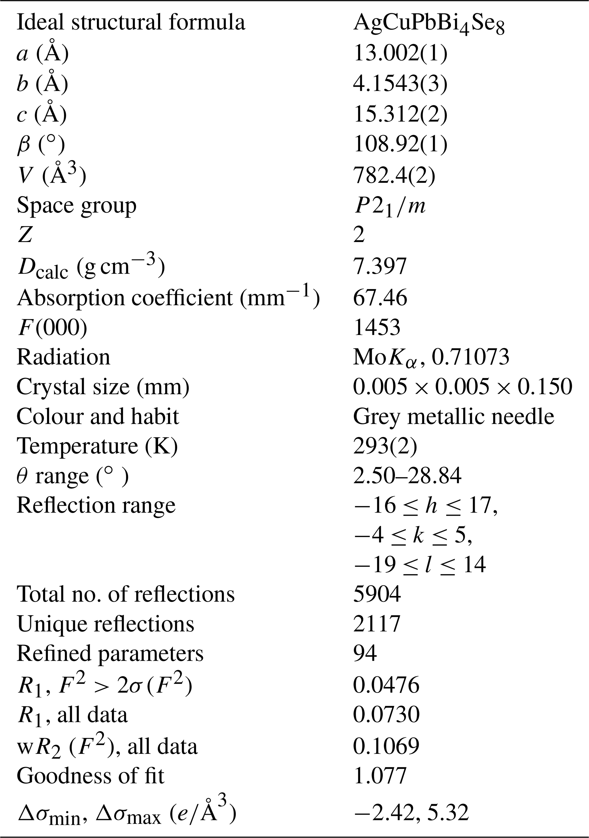
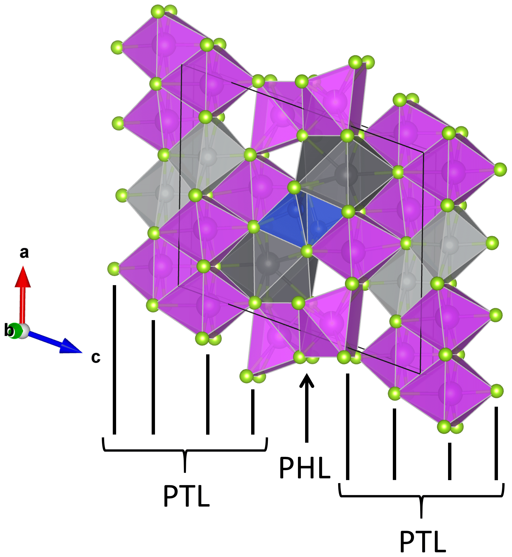
Figure 2The crystal structure of luxembourgite viewed approximately perpendicular to the b axis. Se atoms are yellow, Bi polyhedra are purple, Cu tetrahedra are blue, Ag polyhedra are light grey, and Pb polyhedra are dark grey. Vertical black lines indicate the directions of pseudotetragonal layers (PTLs) and pseudohexagonal layers (PHLs) constituted by Se atoms.
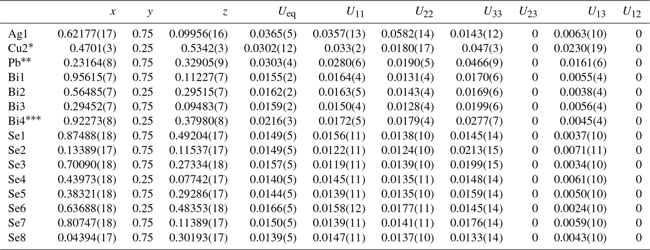
The crystal structure of luxembourgite (Fig. 2; Table 4) was refined in space group P21∕m. Scattering curves for neutral atoms, together with anomalous dispersion corrections, were taken from the International Tables for X-ray Crystallography, Vol. C (Wilson, 1992). In the final refinement cycle, all atoms were refined anisotropically, leading to the R1 value 0.0476. The site occupancy factors were refined for the Cu2, Pb, and Bi4 sites and were constrained to full occupancy for the Ag1, Bi1, Bi2, and Bi3 sites (Table 4). A high residual electron density of 5.32e∕Å3 (Table 3) was observed at 0.94 Å from Bi4, and this atom shows a higher isotropic displacement parameter than other Bi atoms (Table 4). A double occupancy by Bi and Pb was therefore tested for this site, but this model did not improve the R1 value or the displacement parameter of Bi4. Difference Fourier maps indicated an electron density at 0.385 Å from Ag1, so a split-atom model was also tested. The low occupancy factor of the second component of the split Ag atom, 0.015, indicated that this split-atom model was not necessary here.
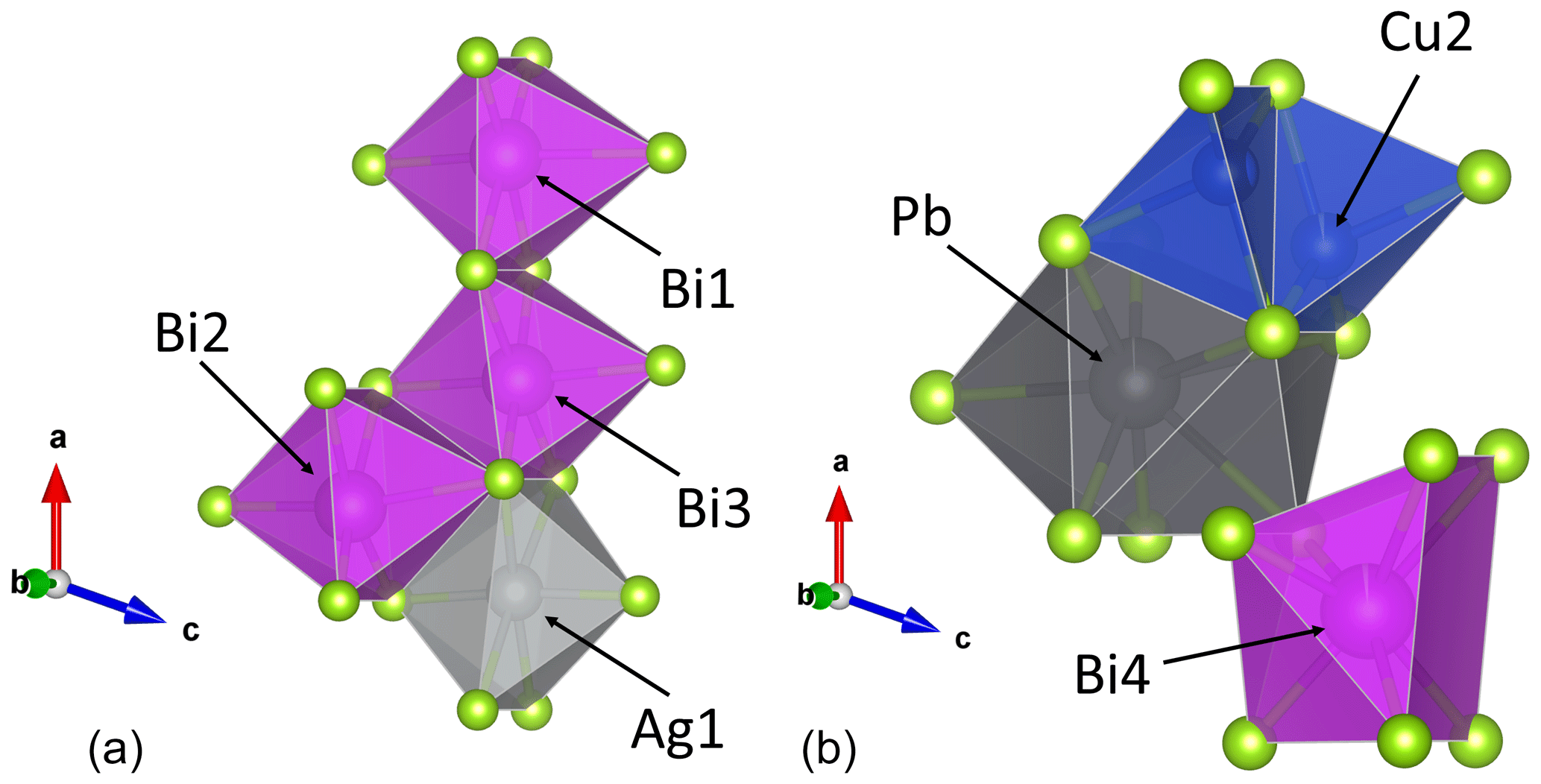
Figure 3Morphologies of the coordination polyhedra in luxembourgite. (a) The Bi1, Bi2, Bi3, and Ag1 octahedra occur in the PbS-like pseudotetragonal layers. (b) The Pb [8], Bi4 [7], and Cu2 [3 + 1] sites occur between the pseudotetragonal and pseudohexagonal anionic layers.
Table 4Atom coordinates and anisotropic displacement parameters (Å2) for luxembourgite from Bivels, Luxembourg.
Occupancies: 0.965(10) Cu; 0.989(4) Pb; 0.977(4) Bi.
The crystal structure of luxembourgite is similar to those of litochlebite (Sejkora et al., 2011) and watkinsonite (Topa et al., 2010), and is based on the stacking of two types of anionic layers: a pseudotetragonal layer four anions thick and a pseudohexagonal layer one anion thick (Fig. 2). In the pseudotetragonal layers the Bi1, Bi2, Bi3, and Ag1 atoms are localised and are each coordinated by six Se anions to form weakly distorted octahedra (Table 5, Fig. 3a). The topology of this layer is identical to that observed in galena, thus explaining why some authors describe the pseudotetragonal layer as a “PbS-like slab” (Topa et al., 2006). The Cu2 atoms are located very close to the pseudohexagonal layer, producing distorted Cu2−Se4 tetrahedra with a [3+1] coordination (Table 5, Fig. 3b). Finally, the Pb and Bi4 atoms are located between the pseudotetragonal and pseudohexagonal slabs, forming coordination polyhedra with rather complex morphologies. Pb is coordinated by five Se anions from the pseudotetragonal layer in a pyramidal arrangement, and by three Se from the pseudohexagonal layer in a triangular arrangement, producing a complex 8-fold polyhedron resembling a pentagonal bipyramid with one supplementary square face (Table 5, Fig. 3b). Such very distorted seven-coordinated site are very typical for Pb, a cation with lone-electron pairs (Theye et al., 2010; Krivovichev and Burns, 2000). Bi4 is coordinated by four Se anions from the pseudotetragonal layer and by three Se anions from the pseudohexagonal layer, leading to a 7-fold polyhedron that can be described as a pyramid with its square face adjusted on a trigonal prism (Table 5, Fig. 3b).
Table 5Selected interatomic distances (Å) for luxembourgite from Bivels, Luxembourg.
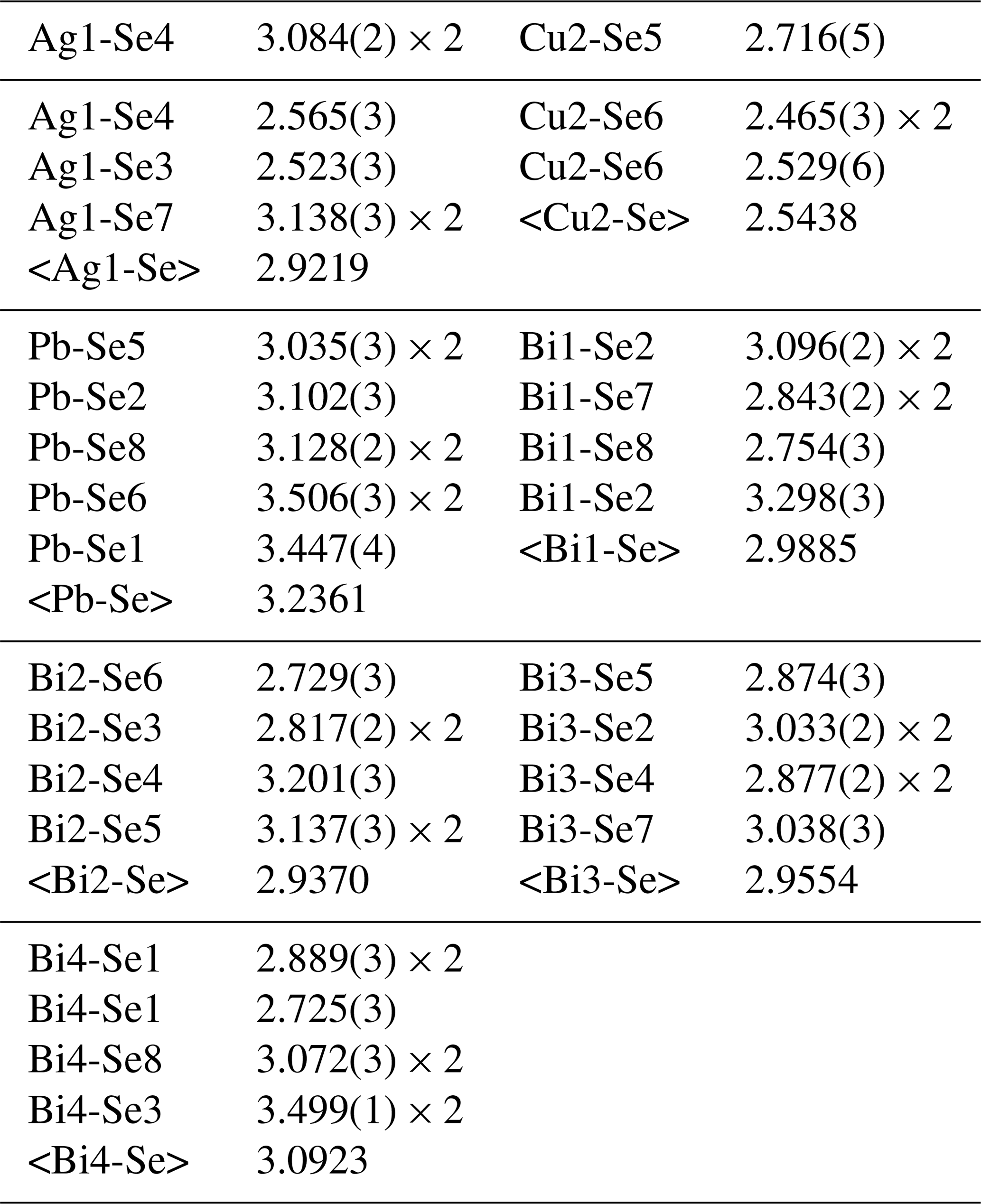
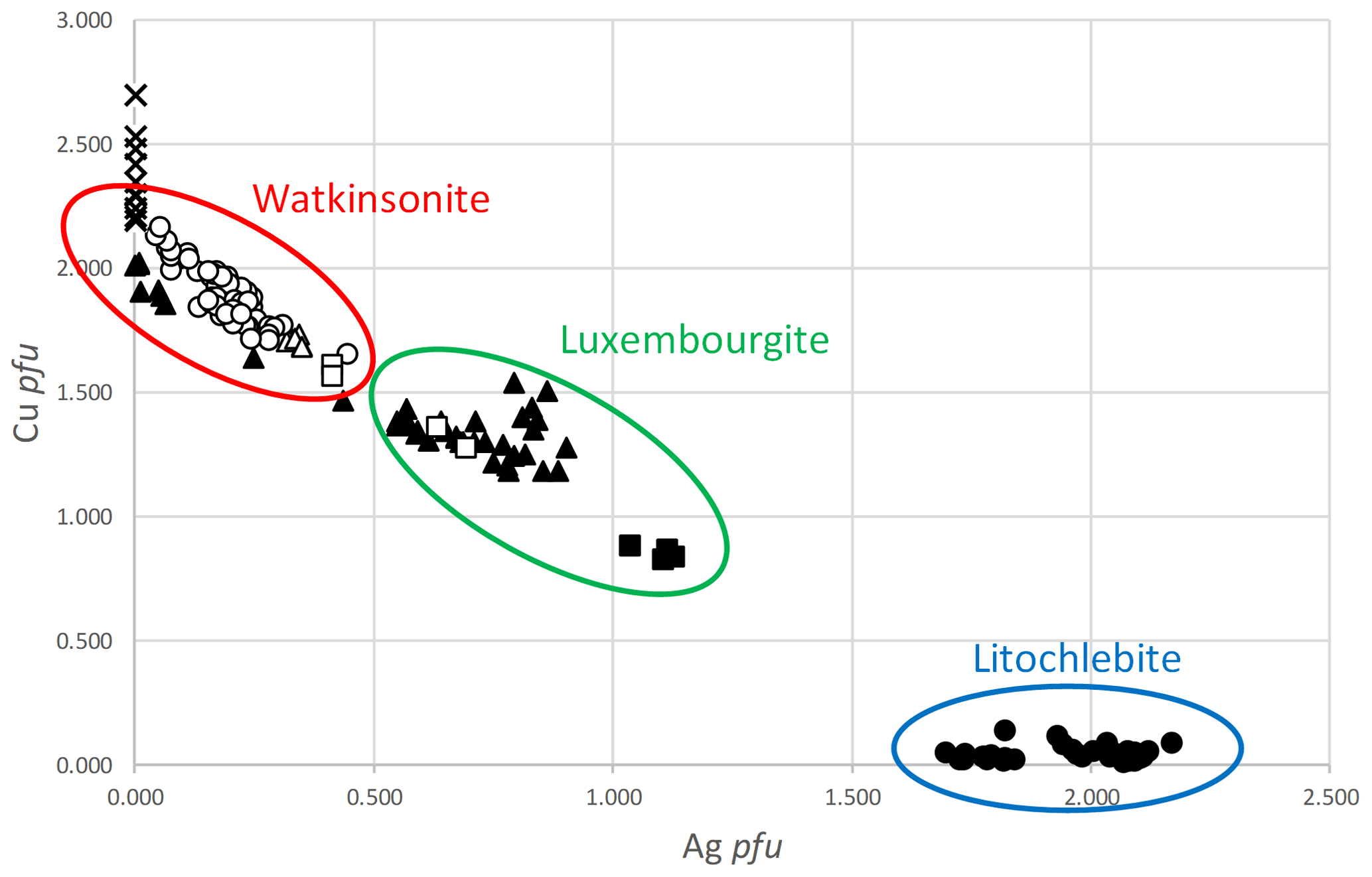
Figure 4Ag ∕ Cu plot, showing the compositions of luxembourgite, watkinsonite, and litochlebite. Crosses represent watkinsonite from Otish Mountains (Johan et al., 1987). Black triangles represent watkinsonite–luxembourgite from Zálesí (Sejkora, unpublished data). White triangles represent watkinsonite from Zálesí (Sejkora et al., 2012). Black circles represent a litochlebite holotype from Zálesí (Sejkora et al., 2011). White circles represent watkinsonite from Zálesí (Topa et al., 2010). Black squares represent luxembourgite from Bivels (this paper). White squares represent watkinsonite–luxembourgite from Niederschlema (Förster et al., 2005).
Site occupancy factors (Table 4) indicate that the Ag1, Bi1, Bi2, and Bi3 sites are fully occupied, while the Cu2 site contains 0.965(10) Cu, the Pb site contains 0.989(4) Pb, and the Bi4 site contains 0.977(4) Bi. Bond distances (Table 5) were used to calculate bond valence sums, according to the empirical relationship established by Brown and Altermatt (1985) and Brese and O'Keeffe (1991). These bond valence sums are 2.08 for Pb, 1.33 for Ag1, 1.00 for Cu2, and between 3.08 and 3.25 for Bi atoms, thus confirming the cationic distributions.
Luxembourgite is isostructural with watkinsonite, Cu2PbBi4Se8, and is closely related to litochlebite, Ag2PbBi4Se8. It also shows similarities with berryite, Cu3Ag2Pb3Bi7S16, a sulfide showing PbS-like slabs containing four Bi and two Ag sites, as well as hexagonal slabs containing Cu atoms coordinated by three S atoms in triangular environments (Topa et al., 2006). In this structure, Pb sites, as well as two Bi sites, occur on the surface of the PbS-like slabs, as observed in the structure of luxembourgite (see above). Table 6 gives a comparison between the properties of luxembourgite, watkinsonite, and litochlebite. In mineralogical classification systems, luxembourgite has a Strunz number 02.HB.20 and a Dana number 3.7.21.
Table 6Comparison of the physical properties of luxembourgite, watkinsonite, and litochlebite.

References: (a) Johan et al. (1987); (b) Topa et al. (2010); (c) Sejkora et al. (2011).
As underlined by Sejkora et al. (2011), the crystal structures of watkinsonite and litochlebite show significant differences, mainly concerning the Ag ∕ Cu crystallographic sites. In watkinsonite, Cu occurs in both octahedral (Cu1) and tetrahedral (Cu2) coordinations, but in litochlebite, all Ag sites occur in octahedral environments. The Ag positions in litochlebite are split on two very close positions, leading to Ag1a–Ag1b and Ag2a–Ag2b pairs of sites, as well as the Cu1 site of watkinsonite, which is split into a Cu1–Ag pair of sites. In luxembourgite, the situation is more ordered, with Ag occurring on the octahedral Ag1 site and Cu on the tetrahedral Cu2 site, without any splitting of atoms. In watkinsonite, Topa et al. (2010) already observed the presence of 0.32 Ag on the Cu1/Ag site; in luxembourgite, this site is fully occupied by Ag (Table 4).
Chemical analyses from the literature (Fig. 4) indicate that a complete solid solution exists between watkinsonite and luxembourgite, but the replacement of Cu by Ag on the Cu2 site is more difficult, certainly due to the small size of this site in watkinsonite and luxembourgite (average M-O bond lengths = 2.544 Å in luxembourgite and 2.730 Å in litochlebite). Consequently, the solid solution between luxembourgite and litochlebite is very limited, due to the significant structural changes between these minerals, and luxembourgite cannot be considered an intermediate phase between litochlebite and watkinsonite. Luxembourgite is the analogue of watkinsonite, with Ag and Cu ordered on the Ag1 and Cu2 sites, respectively.
All data are available upon request to Frédéric Hatert.
The supplement related to this article is available online at: https://doi.org/10.5194/ejm-32-449-2020-supplement.
SP discovered the sample and realised a preliminary EDS analysis. FH performed the single-crystal structure determination with YB, obtained a powder pattern, and wrote the IMA-CNMNC proposal and the final manuscript. PV analysed the species by electron microprobe, and JS indexed the X-ray powder pattern and gave unpublished electron-microprobe analyses of similar samples.
The authors declare that they have no conflict of interest.
The authors are grateful to Baptiste Burnet, Scientific Collaborator of the Natural History Museum of Luxembourg, who collected the luxembourgite samples. The editor Sergey Krivovichev, as well as two anonymous reviewers, are acknowledged for their constructive comments.
This paper was edited by Luca Bindi and reviewed by two anonymous referees.
Beugnies, A.: Le métamorphisme de l'aire anticlinale de l'Ardenne, Hercynia, II, 17–33, 1986.
Brese, N. E. and O'Keeffe, M.: Bond-valence parameters for solids, Acta Cryst., B47, 192–197, 1991.
Brown, I. D. and Altermatt, D.: Bond-valence parameters obtained from a systematic analysis of the Inorganic Crystal Structure Database, Acta Cryst., B41, 244–247, 1985.
Burnham, C. W.: Lattice constant refinement, Carnegie Inst. Washington Yearbook, 61, 132–135, 1962.
Förster, H.-J., Tischendorf, G., and Rhede, D.: Mineralogy of the Niederschlema-Alberoda U-Se-polymetallic deposit, Erzgebirge, Germany. V. Watkinsonite, nevskite, bohdanowiczite and other bismuth minerals, Can. Mineral., 43, 899–908, 2005.
Hatert, F. and Theye, T.: Zeolites, prehnite, and pumpellyite from Bertrix, Belgian Ardennes, Geol. Belgica, 8, 33–42, 2005.
Johan, Z., Picot, P., and Ruhlmann, F.: The ore mineralogy of the Otish Mountains uranium deposit, Québec: skipenite, Bi2Se2Te, and watkinsonite, Cu2PbBi4(Se,S)8, two new mineral species, Can. Mineral., 25, 625–638, 1987.
Krivovichev, S. V. and Burns, P. C.: Crystal chemistry of basic lead carbonates. I. Crystal structure of synthetic shannonite, Pb2O(CO3), Mineral. Mag., 64, 1063–1068, 2000.
Oxford Diffraction: CrysAlis CCD and CrysAlis RED, version 1.71, Oxford Diffraction, Oxford, England, 2007.
Philippo, S. and Hanson, A.: La minéralisation en antimoine de Goesdorf, Ferrantia, 49, 111–146, 2007.
Philippo, S., Hoffmann, I., Faber, A., Heinen, G., Schoellen, J., Schroeder, N., Blom, L., and Bornain, S.: La minéralisation en cuivre de Stolzembourg, Ferrantia, 49, 7–110, 2007.
Philippo, S. and Hatert, F.: La minéralisation en antimoine de Goesdorf, Ferrantia, 77, 7–58, 2018.
Sejkora, J., Makovicky, E., Topa, D., Putz, H., Zagler, G., and Plášil, J.: Litochlebite, Ag2PbBi4Se8, a new selenide mineral from Zálesí, Czech Republic: Description and crystal structure, Can. Mineral., 49, 639–560, 2011.
Sejkora, J., Plášil, J., Litochleb, J., Škácha, P., and Pavlícek, A.R: Association of selenides with macroscopic umangite from the abandoned uranium deposit of Zálesi in the Rychlebské Mountains (Czech Republic), Bull. Mineral. Petrol., 20, 187–196, 2012.
Theye, T. and Fransolet, A. M.: Amphibolitfazielle Metamorphite im Rhenoherzynikum der Ardennen, Ber. Deutschen Mineral. Gesell., Bh. z. Eur. J. Mineral., 5, p. 255, 1993.
Theye, T., Hatert, F., Ockenga, E., Bertoldi, C., and Lathe, C.: Gramaccioliite-(Y): paragenesis, chemical, structural, thermal expansion, and compressibility data on a new occurrence from Samos Island, Greece, Eur. J. Mineral., 22, 443–452, 2010.
Topa, D., Makovicky, E., Putz, H., and Mumme, W. G.: The crystal structure of berryite, Cu3Ag2Pb3Bi7S16, Can. Mineral., 44, 465–480, 2006.
Topa, D., Makovicky, E., Sejkora, J., and Dittrich, H.: The crystal structure of watkinsonite, Cu2PbBi4Se8, from the Zálesí uranium deposit, Czech Republic, Can. Mineral., 48, 1109–1118, 2010.
Wilson, A. J. C.: International Tables for X-ray Crystallography, Vol. C, Kluwer Academic Press, London, 883 pp., 1992.
Yvon, K., Jeitschko, W., and Parthé, E.: Lazy Pulverix, a computer program for calculation X-ray and neutron diffraction powder patterns, J. Appl. Cryst., 10, 73–74, 1977.